Research

Distilling the Data

By Michael Lovett, Ph.D.

The burgeoning field of bioinformatics allows the Hearing Restoration Project to analyze and compare large genomics datasets and identify the best genes for more testing. This sophisticated data analysis will help speed the way toward a cure for hearing loss and tinnitus.


Since its launch in 2011, the Hearing Restoration Project (HRP) is focused on identifying new therapies that will restore inner ear hair cell function, and hence hearing. Within the consortium, smaller research groups engage in separate projects over the course of the year, to move the science along more quickly.

Over the past decade my group, and the group led by my collaborator Mark Warchol, Ph.D., have worked to identify genes that are potential targets for drug development or for gene therapies to cure hearing loss. Our approach has been to determine the exact mechanisms that some vertebrates—in our case, birds—use to regenerate their hair cells and thus spontaneously restore their hearing. We have been comparing this genetic “tool kit” with the mechanisms that mammals normally use to make hair cells.

Unlike birds, mammals cannot regenerate adult hair cells when they are damaged, which is a leading cause of human hearing and balance disorders. Our working hypothesis is that birds have regeneration mechanisms that mammals are missing—or that mammals have developed a repressive mechanism that prevents hair cell regeneration.

In either case, our strategy has been to get a detailed picture of what transpires during hair cell regeneration in birds by using cutting-edge technologies developed during the Human Genome Project (the international research collaboration whose goal was the complete mapping of all the nuclear DNA in humans). These next-generation (NextGen) DNA sequencing methods have allowed us to accurately measure changes in every single gene as chick sensory hair cells regenerate.

The good news is that this gives us, for the first time, an exquisitely detailed and accurate description of all of the genes that are potential players in the process. The bad news is that this is an enormous amount of information; thousands of genes change over the course of seven days of regeneration.

Some of these will be the crucially important—and possibly game-changing—genes that we want to explore in potential therapies, but most will be downstream effects of those upstream formative events. The challenge is to correctly identify the important causative needles in the haystack of later consequences.

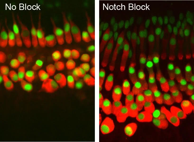
We already know some important genetic players, but we are still far from understanding the genetic wiring of hair cell development or regeneration. For example, after decades of basic research, we know that certain signaling pathways, such as those termed Notch and Wnt, are important in specifying how hair cells develop. These chemical signaling pathways are made of multiple protein molecules, each of which is encoded by a single gene.

However, the Notch and Wnt pathways together comprise fewer than 100 genes and, despite being intensively studied for years, we do not completely understand every nuance of how they fit together.

It also may seem surprising that—more than a decade after the completion of the Human Genome Project and projects sequencing mouse, chick, and many other species’ genomic DNA—we still do not know the exact functions of many of the roughly 20,000 genes, mostly shared, that are found in each organism. This is partly because teasing out all of their interactions and biochemical properties is a painstaking process, and some of the genes exert subtly different effects in different organs. It is also because the genetic wiring diagram in different cells is a lot more complicated than a simple set of “on/off” switches.

All of this sounds a bit dire. Fortunately, we do have some tools for filtering the data deluge into groups of genes that are more likely to be top candidates. The first is to extract all of the information on “known” pathways, such as the Notch and Wnt mentioned earlier. That is relatively trivial and can be accomplished by someone reasonably well versed in Microsoft Excel.

That leaves us with the vast “unknown” world. Analyzing this requires computational, mathematical, and statistical methods that are collectively called bioinformatics. This burgeoning field has been in existence for a couple of decades and covers the computational analysis of very large datasets in all its forms. For example, we routinely use well-established bioinformatic methods to assemble and identify all of the gene sequences from our NextGen DNA sequence reads. These tasks would take many years if done by hand, but a matter of hours by computational methods.

In the case of our hair cell regeneration data, our major bioinformatic task is to identify the best genes for further experimental testing. One method is to computationally search the vast biological literature to see if any of them can be connected into new networks or pathways. There are now numerous software tools for conducting these types of searches. However, this really is not very helpful when searching through several thousand genes at once. The data must be filtered another way to be more useful.

We have used statistical pattern matching tools called self-organizing maps to analyze all of our data across every time point of hair cell regeneration. In this way we can detect genes that show similar patterns of changes and then drill down deeper into whether these genes are connected. This has provided us with an interesting “hit list” of genes that have strong supporting evidence of being good candidates for follow-up.

An additional approach is to compare our chick data to other datasets that the HRP consortium is collecting. The logic here is that we expect key genetic components to be shared across species. For example, we now know a great deal about what genes are used in zebrafish hair cell regeneration and the genes that specify mouse hair cells during normal development. We can conduct computational comparisons across these big datasets to identify what is similar and what is different. Again, this has yielded a small and interesting collection of genes that is being experimentally tested. 

Our final strategy has been to extract classes of genes that act as important switches in development. These transcription factors control other genetic circuits. We have identified all of these that change during chick hair cell regeneration. As a consortium the HRP now has a collection of about 200 very good candidate genes for follow-up. However, software and high-speed computation are not going to do it all for us. We still need biologists to ask and answer the important questions and to direct the correct bioinformatics comparisons.

Hair cell regeneration is a plausible goal for the treatment of hearing and balance disorders. The question is not if we will regenerate hair cells in humans, but when. Your financial support will help to ensure we can continue this vital research and find a cure in our lifetime! Please help us accelerate the pace of hearing and balance research and donate today. Your HELP is OUR hope!

If you have any questions about this research or our progress toward a cure for hearing loss and tinnitus, please contact Hearing Health Foundation at info@hhf.org.

Michael Lovett, Ph.D., is a professor at the National Lung & Heart Institute in London and the chair in systems biology at Imperial College London.

Print Friendly and PDF

BLOG ARCHIVE

Getting a Hearing Test May Be Good for Your Memory

By the Better Hearing Institute

If you want to help your memory and cognitive performance, you may want to get a hearing test and treat hearing loss. In response to a growing body of research that shows a link between unaddressed hearing loss and cognitive function, the Hearing Health Foundation (HHF) and the Better Hearing Institute (BHI) are encouraging people to get their hearing checked by a healthcare professional in recognition of World Alzheimer’s Month in September.


According to Brandeis University Professor of Neuroscience, Dr. Arthur Wingfield, who has been studying cognitive aging and the relationship between memory and hearing acuity for many years, effortful listening due to unaddressed hearing loss is associated with increased stress and poorer performance on memory tests.
 
His research shows that even when people with unaddressed hearing loss perceive the words that are being spoken, their ability to remember the information suffers—likely because of the draw on their cognitive resources that might otherwise be used to store what has been heard in memory. This is especially true for the comprehension of quick, informationally complex speech that is part of everyday life.
 
“Even if you have just a mild hearing loss that is not being treated, cognitive load increases significantly,” Wingfield said. “You have to put in so much effort just to perceive and understand what is being said that you divert resources away from storing what you have heard into your memory.”
 
How hearing loss affects cognitive function
Our ears and auditory system bring sound to the brain. But we actually “hear” with our brain, not with our ears.
 
According to Wingfield, unaddressed hearing loss not only affects the listener’s ability to perceive the sound accurately, but it also affects higher-level cognitive function. Specifically, it interferes with the listener’s ability to accurately process the auditory information and make sense of it.
 
In one study, Wingfield and his co-investigators found that older adults with mild to moderate hearing loss performed poorer on cognitive tests of memory than those of the same age who had good hearing.
 
In another study, Wingfield and colleagues at the University of Pennsylvania and Washington University in St. Louis used MRI to look at the effect that hearing loss has on both brain activity and structure. The study found that people with poorer hearing had less gray matter in the auditory cortex, a region of the brain that is necessary to support speech comprehension.
 
Wingfield has suggested the possibility that the participant’s hearing loss had a causal role. He and his co-investigators hypothesize that when the sensory stimulation is reduced due to hearing loss, corresponding areas of the brain reorganize their activity as a result.
 
“The sharpness of an individual’s hearing has cascading consequences for various aspects of cognitive function,” said Wingfield. “We’re only just beginning to understand how far-reaching these consequences are.”
 
As people move through middle age and their later years, Wingfield suggested, it is reasonable for them to get their hearing tested annually. If there is a hearing loss, it is best to take it seriously and treat it.
 
Hearing loss and dementia

A number of studies have come to light over the last few years showing a link between hearing loss and dementia.  Specifically, a pair of studies out of Johns Hopkins found that hearing loss is associated with accelerated cognitive decline in older adults and that seniors with hearing loss are significantly more likely to develop dementia over time than those who retain their hearing. 
 
A third Johns Hopkins study revealed a link between hearing loss and accelerated brain tissue loss. The researchers found that for older adults with hearing loss, brain tissue loss happens faster than it does for those with normal hearing.
 
Some experts believe that interventions, like hearing aids, could potentially delay or prevent dementia. Research is ongoing
 
Staying connected

A number of studies indicate that maintaining strong social connections and keeping mentally active as we age might lower the risk of cognitive decline and Alzheimer's disease, according to the Alzheimer’s Association website.
 
Interestingly, BHI research shows that people with hearing difficulty who use hearing aids are more likely to have a strong support network of family and friends, feel engaged in life, and meet up with friends to socialize. They even say that using hearing aids has a positive effect on their relationships.

For more information about hearing health and finding a healthcare professional, please visit: http://hearinghealthfoundation.org/find-a-hearing-health-professional.

The content for this blog post originated in a press release issued by The Better Hearing Institute on September 8, 2015.

Print Friendly and PDF

BLOG ARCHIVE

Emerging Research Grants: 2016 Application Period is Now Open

By Laura Friedman

We are excited to inform you that the application period for Hearing Health Foundation's 2016 Emerging Research Grants program is now open. 

Through ERG, HHF, the largest non-profit funder of hearing and balance research, awards emerging researchers grants to conduct novel investigations of auditory and vestibular function and dysfunction. HHF wishes to stimulate research that leads to a continuing and independently fundable line of research. Only research proposals in the topic areas listed in our announcement, including basic, translational, and applied clinical research, will be considered:

We encourage you to review our announcement and Policy on Emerging Research Grants. If you are eligible to apply for this program, please make note of the deadlines below and review the instructions for submitting a letter of intent (LOI).

2016 HHF-ERG Timeline for Applicants:

  • LOI deadline: October 26, 2015 by 5pm ET

  • Full Application opens: Early November, 2015

  • Full Application deadline: December 7, 2015 by 5pm ET

If you have any questions about the ERG program and process, please contact us at grants@hhf.org

Please forward and share this information with your colleagues. 

Print Friendly and PDF

BLOG ARCHIVE

Orchestrating Hair Cell Regeneration

By the Stowers Institute for Medical Research

The older we get, the less likely we are to hear well, as our inner ear sensory hair cells succumb to age or injury. Intriguingly, humans are one-upped by fish here. Similar hair cells in a fish sensory system that dots their bodies and forms the lateral line, by which they discern water movement, are readily regenerated if damage or death occurs.

A neuromast sensory structure (green) of the zebrafish lateral line, which helps the fish detect water movement, is shown among surrounding cells (cell nuclei in red).Credit: Piotrowski Lab, Stowers Institute for Medical Research

A neuromast sensory structure (green) of the zebrafish lateral line, which helps the fish detect water movement, is shown among surrounding cells (cell nuclei in red).

Credit: Piotrowski Lab, Stowers Institute for Medical Research

A new study in the July 16 online and August 10 print issue of Developmental Cell, from Stowers Institute for Medical Research Associate Investigator Tatjana Piotrowski, Ph.D., zeros in on an important component of this secret weapon in fish: the support cells that surround centrally-located hair cells in each garlic-shaped sensory organ, or neuromast. “We’ve known for some time that fish hair cells regenerate from support cells,” Piotrowski explains, “but it hasn’t been clear if all support cells are capable of this feat, or if subpopulations exist, each with different fates.”

While mammals also have support cells, they unfortunately do not respond to hair cell death in the same way. So understanding how zebrafish support cells respond to hair cell loss may provide insight into how mammalian support cells might be coaxed into regenerating hair cells as well. Zebrafish are particularly amenable to studies of regeneration because transparent embryos and larvae render developmental processes visible and experimentally accessible.

Piotrowski and her team treated zebrafish larvae with the antibiotic neomycin, which kills hair cells, then monitored support cell proliferation in regenerating neuromasts for three days using time-lapse movies. “These single cell lineage analyses were tremendously time-consuming but very informative,” Piotrowski notes. The study’s lead author, Andrés Romero-Carvajal, Ph.D., previously a predoctoral researcher at the Stowers Institute, carefully kept track of every individual support cell’s location and behavior across different time-lapse frames.

The researchers determined that approximately half of the dividing support cells differentiated into hair cells, while the rest self-renewed. Self-renewal is an equally important fate, Piotrowski points out, because it ensures maintenance of a reserve force that, if necessary, can spring into regenerative action. The researchers also observed that lineage fate of support cells hinged on where they were located in the neuromast, as self-renewing cells were found clustered at opposite poles while differentiating cells were distributed in a random, circular pattern close to the center. 

Such distinct support cell locations were “strongly indicative of differences in gene expression”, Piotrowski says, so the team turned its attention to exploring some of the genes and signaling pathways involved. A study of gene expression patterns showed that members of the Notch and Wnt pathways were expressed in different parts of the neuromast, specifically the Notch members in the center and the Wnt members at the poles. To determine if and how these two pathways regulate each other, the researchers used an inhibitor to turn off Notch signaling in neuromasts. This halt in Notch activity mimics the halt known to occur immediately after neomycin-induced hair cell death. After inhibitor treatment, they saw transient upregulation of Wnt ligands in the neuromast center, along with support cell proliferation. The majority of the proliferating cells became hair cells.

“We found that Notch directly suppresses differentiation (of support cells into hair cells), and indirectly inhibits proliferation by keeping Wnt in check,” Piotrowski explains. “Previously, others thought perhaps it was Wnt that had to be downregulated, to initiate regeneration. However, our data support the loss of Notch signaling as a more likely trigger.” Essentially, the process of restoring injured or dead hair cells in neuromasts is jump-started by the transient suppression of Notch, while its eventual reactivation restores the balance, ensuring that not all support cells answer the call to regenerate through proliferation and differentiation.

Piotrowski’s research is partially supported by the Hearing Health Foundation through its Hearing Restoration Project (HRP), which emphasizes collaborations across multiple institutions to develop new therapies for hearing loss. By continuing to illuminate the intricacies of hair cell regeneration in zebrafish, she and her team are providing other HRP scientists with candidate genes and molecular pathways to probe in other models such as chicken and mice, with the goal of providing insight that could someday make human inner ear hair cells readily replaceable.

The study was also funded by the Stowers Institute and the National Institute on Deafness and Other Communication Disorders of the National Institutes of Health (award RC1DC010631). The content is solely the responsibility of the authors and does not necessarily represent the official views of the NIH.

Other Institute contributors include Joaquín Navajas Acedo; Linjia Jiang, Ph.D.; Agnė Kozlovskaja-Gumbrienė; Richard Alexander; and Hua Li, Ph.D.

Lay summary of findings

Hair cells in sensory structures called neuromasts, which form the sensory system fish use to orient themselves in water, are similar to mammalian inner ear hair cells responsible for our sense of hearing. Unlike the latter, however, they are constantly replaced after damage or death. In the current issue of Developmental Cell, Stowers Associate Investigator Tatjana Piotrowski, Ph.D., and members of her lab closely examine, in zebrafish, the support cells from which hair cells regenerate. By tracking individual support cells during neuromast regeneration, first author Andrés Romero-Carvajal, Ph.D., shows that approximately half become hair cells, while the rest self-renew as support cells. These lineage decisions are coordinated by interactions between the Notch and Wnt signaling pathways and are location-specific, as differentiation into hair cells occurs toward the center of neuromasts and self-renewal occurs at opposite poles of the structures. Piotrowski hopes her lab’s findings in zebrafish may be extrapolated to mammals someday, to help provide basic insight needed to progress towards the ultimate goal of regenerating human inner ear hair cells.

About the Stowers Institute for Medical Research

The Stowers Institute for Medical Research is a non-profit, basic biomedical research organization dedicated to improving human health by studying the fundamental processes of life. Jim Stowers, founder of American Century Investments, and his wife, Virginia, opened the Institute in 2000. Since then, the Institute has spent over one billion dollars in pursuit of its mission.

Currently, the Institute is home to almost 550 researchers and support personnel; over 20 independent research programs; and more than a dozen technology-development and core facilities.

The above post is reprinted, with permission, from materials provided by Stowers Institute for Medical Research.

Your financial support will help to ensure we can continue this vital research and find a cure in our lifetime! Please help us accelerate the pace of hearing and balance research and donate today. Your HELP is OUR hope!

If you have any questions about this research or our progress toward a cure for hearing loss and tinnitus, please contact Hearing Health Foundation at info@hhf.org.

Print Friendly and PDF

BLOG ARCHIVE

2015 Emerging Research Grants Approved!

By Laura Friedman

Hearing Health Foundation is excited to announce that the 2015 Emerging Research Grants (ERG) have been approved by our Board of Directors, after a rigorous scientific review process. The areas that we are funding for the 2015 cycle are:

  • Central Auditory Processing Disorder (CAPD): Four grants were awarded for innovative research that will increase our understanding of the causes, diagnosis, and treatment of central auditory processing disorder, an umbrella term for a variety of disorders that affect the way the brain processes auditory information. All four of our CAPD grantees are General Grand Chapter Royal Arch Masons International award recipients.

  • Hyperacusis: Two grants were awarded that is focused on innovative research (e.g., animal models, brain imaging, biomarkers, electrophysiology) that will increase our understanding of the mechanisms, causes, diagnosis, and treatments of hyperacusis and severe forms of loudness intolerance. Research that explores distinctions between hyperacusis and tinnitus is of special interest. Both of our Hyperacusis grants were funded by Hyperacuis Research.

  • Ménière’s Disease: Two grants were awarded for innovative research that will increase our understanding of the inner ear and balance disorder Ménière’s disease. One of the grants is funded by The Estate of Howard F. Schum and the other is funded by William Randolph Hearst Foundation through their William Randolph Hearst Endowed Otologic Fellowship.

  • Tinnitus: Two grants were awarded for innovative research that will increase our understanding of the mechanisms, causes, diagnosis, and treatment of tinnitus. One of the grants is funded by the Les Paul Foundation and the other grantee is the recipient of The Todd M. Bader Research Grant of The Barbara Epstein Foundation, Inc.

To learn more about our 2015 ERG grantees and their research proposals and goals, please visit: http://hearinghealthfoundation.org/2015_researchers

Hearing Health Foundation is also currently planning for our 2016 ERG grant cycle. If you're interested in naming a research grant in any discipline within the hearing and balance space, please contact development@hhf.org.

Print Friendly and PDF

BLOG ARCHIVE

Rare Gene Cariant Associated with Middle Ear Infections

By Baylor College of Medicine

Many parents have heard the night-time cry of “my ear hurts.” For some children, this might happen frequently beginning in infancy and even persist into adulthood. An international consortium led by those at Baylor College of Medicine may have taken the first step on the road to understanding why only some people get frequent painful or chronic middle ear infections. The culprit may be rare genetic variants in a gene called A2ML1.

A report on their work appears online in the journal Nature Genetics.


In studies led by Dr. Regie Lyn P. Santos-Cortez, assistant professor of molecular and human genetics at Baylor, researchers looked for a genetic component to the disorder. Santos-Cortez is not only a genetics researcher, she was also trained as an otolaryngologist in her native Philippines, and she knows the toll such infections take.

After graduation, she went on a medical missionary trip to an indigenous Filipino population in one area of the country where most of the people were related.

There she created a family tree or pedigree that identified, among other things, who within the same community suffered from recurrent ear infections and who did not.

“The pedigree was huge,” she said. “It was several pages long and wide.”

Everyone had similar socioeconomic status, swam in the same sea water, were or had been mostly breastfed, ate the same food, and had the same exposure to cigarette smoke, which made an environmental factor an unlikely cause.

Luckily, next-generation sequencing that allowed her to determine the genetic sequence of several people in the population was available. Without that technological advance, she said, she did not think they could have identified the gene.

Within the indigenous community, she found that 80 percent of those who carry the variant in the A2ML1 gene developed otitis media. They also found the same gene variant in three otitis-prone children in a group in Galveston, Texas.

So far, they have identified this rare genetic cause for susceptibility to middle ear infections in 37 Filipinos, one Hispanic-American and two European-Americans. It is likely that the variant has been present in the population in the Philippines and in Galveston at least 150 years and may even be the result of a “founder” effect, which suggests one person from outside the population, more likely from Spain, brought the gene variant into the two populations.

Additionally, rare A2ML1 variants were identified in six otitis-prone children who were Hispanic- or European-American, and none of these variants occurred in thousands of individuals without otitis media.

She does not think this is the only gene involved in predisposing children to middle ear infections, but it could be an important one. The protein involved may play a role in the immune system that protects the ear. Perhaps the variant somehow derails the protection the protein should provide.

Another gene called alpha 2-macroglobulin or A2M, which encodes a protein that is found at high levels in very infected ears, is formed in such a way that it can trap proteases, enzymes that can kill infectious microbes but can also damage the mucosa of the middle ear if left unchecked.

Because the protein sequences of A2M and A2ML1 are highly identical, they may have similar or overlapping functions and one might compensate for the other when it is non-functional. An antibiotic drug called bacitracin is used in drop form to treat the problem in Europe. However, because bacitracin dampens the effect of A2M it may not be the best treatment for people who have genetic variants in A2ML1, she said.

“There are many other antibiotic drops on the market,” said Santos-Cortez.

The finding of the variant is a start, she said. She and her colleagues hope to look further into the mechanism by which A2ML1 defects cause otitis media susceptibility.

Others who took part in this work include Xin Wang, Anushree Acharya, Izoduwa Abbe, Biao Li, Gao T. Wang and Suzanne M. Leal, all of Baylor; Charlotte M. Chiong, Ma Rina T Reyes-Quintos, Ma Leah C. Tantoco, Marieflor Cristy Garcia, Erasmo Gonzalo D V Llanes, Patrick John Labra, Teresa Luisa I. Gloria-Cruz, Abner L. Chan, Eva Maria Cutiongco-de la Paz and Generoso T. Abes, all of the University of the Philippines Manila-National Institutes of Health; Arnaud P. Giese, Saima Riazuddin and Zubair M. Ahmed, University of Maryland at Baltimore; Joshua D Smith, Jay Shendure, Michael J. Bamshad and Deborah A. Nickerson, all of the University of Washington at Seattle; E. Kaitlynn Allen and Michele M. Sale of the University of Virginia in Charlottesville; Kathleen A. Daly of the University of Minnesota in Minneapolis; Janak A. Patel and Tasnee Chonmaitree of the University of Texas Medical Branch at Galveston.

Funding for this work came from the Hearing Health Foundation; Action On Hearing Loss and the National Organization for Hearing Research Foundation (to R.L.P.S.-C.); the University of the Philippines Manila–National Institutes of Health (to G.T.A. and R.L.P.S.-C.); and U.S. National Institutes of Health (Grants U54 HG006493 (to D.A.N.), R01 DK084350 (to M.M.S.), R01 DC003166 (to K.A.D.), R01 DC005841 (to T.C.), R01 DC011803 and R01 DC012564 (to S.R. and Z.M.A.), and R01 DC011651 and R01 DC003594 (to S.M.L.).

The above post is reprinted from materials provided by Baylor College of Medicine.

Help us change the course of hearing research and find a cure for hearing loss and tinnitus! Hearing Health Foundation’s “Name a Research Grant” program enables donors to name and fund a specific research grant in their name or in honor or memory of a loved one.

We're currently planning for our 2016 grant cycle. If you're interested in naming a research grant in any discipline within the hearing and balance space, such as Usher Syndrome, hyperacusis, stria, or tinnitus, please contact development@hhf.org

Print Friendly and PDF

BLOG ARCHIVE

Life-Saving Antibiotics Increase Risk of Hearing Loss

By Oregon Health & Science University

Approximately 80% of infants in neonatal intensive care units receive antibiotics known to be toxic to the ear. 

Seeking to stem the tide of permanent hearing loss from the use of life saving antibiotics, researchers at Oregon Health & Science University have found that patients stricken with dangerous bacterial infections are at greater risk of hearing loss than previously recognized. Inflammation from the bacterial infections substantially increased susceptibility to hearing impairment by increasing the uptake of aminoglycoside antibiotics into the inner ear, the researchers report. Their findings are published in online in the journal Science-Translational Medicine.

“Currently, it’s accepted that the price that some patients have to pay for surviving a life-threatening bacterial infection is the loss of their ability to hear. We must swiftly bring to clinics everywhere effective alternatives for treating life-threatening infections that do not sacrifice patients’ ability to hear,” said Peter S. Steyger, Ph.D.*, professor of otolaryngology, head and neck surgery, Oregon Hearing Research Center, Oregon Health & Science University School of Medicine. “Most instances in which patients are treated with aminoglycosides involve infants with life-threatening infections. The costs of this incalculable loss are borne by patients and society. When infants lose their hearing, they begin a long and arduous process to learn to listen and speak. This can interfere with their educational trajectory and psychosocial development, all of which can have a dramatic impact on their future employability, income and quality of life.”

Aminoglycosides, antimicrobials that are indispensable to treating life-threatening bacterial infections, are toxic to the ear. Relied on by physicians to treat meningitis, bacteremia and respiratory infections in cystic fibrosis, aminoglycosides kill the sensory cells in the inner ear that detect sound and motion.

Infants in neonatal intensive care units, or NICUs, are at particular risk. Each year, approximately 80 percent of 600,000 admissions into NICUs in the United States receive aminoglycosides. The rate of hearing loss in NICU graduates is 2 to 4 percent compared with 0.1 to 0.3 percent of full-term births from congenital causes of hearing loss.

When Steyger and colleagues gave healthy mice a low amount of aminoglycoside, the rodents experienced a small degree of hearing loss. If the mice had an inflammation that is typical of the infections treated with aminoglycosides in humans, the mice experienced a vastly greater degree of hearing loss.

The study lays the groundwork for improving the standard of care guidelines for patients receiving aminoglycosides. To shield patients’ hearing, the researchers called for the development of more targeted aminoglycosides and urged clinicians to choose more targeted, non-ototoxic antibiotics or anti-infective drugs to treat patients stricken with severe infections.

Due to their widespread availability and low cost, aminoglycosides are used frequently worldwide. Clinical use of aminoglycosides is limited due to the known risk of acute kidney poisoning and permanent hearing loss, yet are crucial life-savers in cases with potentially fatal infections.

Scientists who contributed to the OHSU study, “Endotoxemia-mediated inflammation potentiates aminoglycoside-induced ototoxicity,” include: Steyger; Ja-won Koo, M.D., Ph.D.; Lourdes Quintanilla-Dieck, M.D.; Meiyan Jiang, Ph.D.; Jianping Liu, M.D., Ph.D.; Zachary D. Urdang, B.S.; Jordan Allensworth, B.S.; Campbell P. Cross, B.A.; and Hongzhe Li, Ph.D.

This research was supported by: National Research Foundation of Korea grant 2011-0010166; Seoul National University Bundang Hospital 03-2011-007 (J.K.W.); National Institute of Deafness and Other Communication Disorders R01 DC004555, R01 DC12588 (P.S.S.), R03 DC011622 (H.L.), and P30 DC005983; and the Department of Otolaryngology at OHSU (L.Q.D.).

*Peter S. Steyger, Ph.D., is a prior Hearing Health Foundation board member and previous head of our Council of Scientific Trustees.

The above post is reprinted from materials provided by Oregon Health & Science University.

Print Friendly and PDF

BLOG ARCHIVE

Ask the Scientist: Gene Therapies and Hearing

By Peter G. Barr-Gillespie, Ph.D.

A DNA double helixNational Human Genome Research Institute

A DNA double helix

National Human Genome Research Institute

Recently, Hearing Health Foundation (HHF) has received several questions regarding the Reuters report on gene therapies for hearing. There are two separate but related topics raised in this article. As the scientific research director of HHF’s Hearing Restoration Project, which since 2011 has been uncovering concrete discoveries toward a biologic cure for hearing loss and tinnitus, I want talk about each individually, and then discuss what I interpret they mean together.


The article first presents the Science Translational Medicine paper from Jeffrey Holt’s lab. This is very much a proof-of-principle report, focused on an animal model and using a time for delivery of the corrected gene that is extremely early in development (equivalent to a 5-to-6-month-old human fetus). It is important to point out that their strategy will only correct one type of genetic hearing loss and genetic hearing loss from mutations in other genes will require related but different strategies. Nevertheless, this is an exciting example of modeling gene therapy in animals, and represents a logical progression toward that goal in humans.

The article then moves on to reference the Novartis trial. For this trial, they are using a similar technical strategy, viral delivery of a gene, but they are targeting people—those who have lost their hearing through non-genetic means, such as noise damage, aging, or infections. The gene they are delivering, known as ATOH1, may stimulate production of new hair cells; it is a gene that is essential for formation of hair cells during development, and in some experimental animal models, delivery of the gene can lead to production of a few hair cells in adult ears.

That said, many people who I have talked to in the field who work with experimental models of hair cell formation using ATOH1, including members of our Hearing Restoration Project consortium, believe that this trial is premature. By and large, the animal models do not support the trial; most suggest that there will be few hair cells formed and little hearing restored. While we can hope for a little bit of hearing recovery, we are concerned about toxic responses to the gene delivery using viruses. Personally, while I think it would be truly fantastic if the Novartis trial works, at this moment in time I don’t think the rewards yet outweigh the considerable risks being imposed on a human (include safety during the procedure and potential side effects afterward).

Still, the Novartis trial will tell us about the safety of viral delivery into the ears of humans, and knowing that is critically important. I think the most likely outcome is that we will learn whether the strategy the Novartis trial used to deliver the gene is safe. Unfortunately, if we don’t see improved hearing, we won’t know why—did the gene not get to the right place, or does it just not work?

Technical aspects of gene delivery are what ties together the Novartis work and the Holt lab work. Both use viruses for delivering genes, and together the results from these and others will let us know, from a procedural standpoint, how we can deliver genes to the ear. I think it is unlikely that delivering just ATOH1 will do the trick of restoring hearing; it may be that we need to deliver other genes or to use drugs to overcome the block we see to making new hair cells.

So while these are exciting reports to hear about, especially that Novartis is actually carrying out a trial in humans, it is still premature to think that this is going to be a viable strategy for restoring hearing. This is why Hearing Health Foundation's Hearing Restoration Project is doing everything possible to accelerate the pace of its research.

Hair cell regeneration is a plausible goal for the treatment of hearing and balance disorders. The question is not if we will regenerate hair cells in humans, but when. Your financial support will help to ensure we can continue this vital research and find a cure in our lifetime! Please help us accelerate the pace of hearing and balance research and donate today. Your HELP is OUR hope!

If you have any questions about this research or our progress toward a cure for hearing loss and tinnitus, please contact Hearing Health Foundation at info@hhf.org.

Print Friendly and PDF

BLOG ARCHIVE

Taking Hair Cell Regeneration Down a Notch

By Andy Groves

Hearing Restoration Project (HRP) consortium scientists, Andy Groves and Stefan Heller had their research published in Frontiers in Cellular Neuroscience on March 31, 2015. Below is a summary of their research:

Sensorineural hearing loss is most commonly caused by the death of hair cells in the organ of Corti, and once lost, mammalian hair cells do not regenerate. In contrast, other vertebrates such as birds can regenerate hair cells by stimulating division and differentiation of neighboring supporting cells.

During the development of the inner ear, newly-born hair cells send signals to their neighbors that instruct them to not become hair cells, but to become supporting cells instead. Hair cells are the mechanosensitive cells of the ear. Supporting cells surround them and as their name implies, physically support them and help regulate some of the properties of hair cells. One of these signals is an evolutionarily ancient pathway, the Notch signaling pathway. We and others have shown that if you block the Notch signaling pathway in the cochlea or balance organs of young mice, the supporting cells no longer get the message to stay as supporting cells, and instead they transform into hair cells. This process also happens during hair cell regeneration in birds - supporting cells transform into hair cells, which then send a Notch signal to their neighbors and prevent too many hair cells from being formed.

These observations suggest that it might be possible to block Notch signaling in mature, deafened animals as a means of getting new hair cells to form. We performed a simple experiment to test this in progressively older and older animals. To our surprise, we found that once mice are more than a week old, blocking Notch signaling has no effect on the cochlea any more, and no new hair cells are made.  We showed that this was due in part to components of the Notch signaling pathway being switched off in the ear as the animals get older. Viewed this way, the Notch signaling pathway can be thought of as a “Scaffold” - it is used to allow the cochlea to be built in the first place, but is then dismantled once the cochlea becomes functional.

What does this mean? It suggests that inhibiting Notch signaling alone is unlikely to be an effective means of hair cell regeneration in mammals. It is possible that other factors will be required, and some HRP members are busy testing these other pathways right now. It will also be of great interest to understand HOW the Notch pathway is dismantled with age, whether we can exploit this in future therapies.

Read more about this research proposal here: http://hearinghealthfoundation.org/hrp-consortium-projects-groves-segil-stone.

This work was supported by Department of Defense Grant DODW81XWH-11-2-004(AKG) and Hearing Restoration Project consortium grants from the Hearing Health Foundation (AKG and SH), NIH grant DC004563 (SH), NIH grant P30DC010363 (SH, JSO), and NIHR01DC014450 (JSO).

Hair cell regeneration is a plausible goal for the treatment of hearing and balance disorders. The question is not if we will regenerate hair cells in humans, but when. Your financial support will help to ensure we can continue this vital research and find a cure in our lifetime! Please help us accelerate the pace of hearing and balance research and donate today. Your HELP is OUR hope!

If you have any questions about this research or our progress toward a cure for hearing loss and tinnitus, please contact Hearing Health Foundation at info@hhf.org.

Print Friendly and PDF

BLOG ARCHIVE

Gene Discoveries May Lead to Regeneration of Cells Needed for Hearing

By Jeffrey Norris

The researchers identified patterns of gene expression that may determine whether the ear’s inner pillar cells can give rise to new hair cells, which are key to hearing.

School of Medicine scientists have discovered biological mechanisms that appear to play a role in the regeneration of cells in the inner ear.

Over a lifetime, these cells often are damaged or die due to oxidative stress, excessive noise exposure or toxic drugs. The accumulated loss can significantly compromise hearing. Nearly one in four people ages 65-74, and half who are 75 or older, are candidates for hearing aids because of disabling hearing loss.

The discoveries could lead to new ways of evaluating, in animal models, experimental drug treatments intended to prevent hearing loss or restore hearing, and might even lead to methods for regenerating vital cells that have been lost, said Stefan Heller, PhD, professor of otolaryngology.

A paper describing the findings, as well as new methods to quickly link changes in cell function during development to molecular changes within cells, was published June 9 in Cell Reports. Heller is the senior author of the paper. Postdoctoral scholars Jöerg Waldhaus, PhD, and Robert Durruthy-Durruthy, PhD, share the lead authorship.

Discoveries by Stefan Heller and his colleagues could lead to new ways of evaluating, in animal models, experimental drug treatments intended to prevent hearing loss or restore hearing. - Steve Fisch

Discoveries by Stefan Heller and his colleagues could lead to new ways of evaluating, in animal models, experimental drug treatments intended to prevent hearing loss or restore hearing. - Steve Fisch

Sound waves striking the eardrum cause vibrations that are transmitted through tiny bones in the middle ear to fluid within the snail-shell-shaped cochlea of the inner ear. Specialized cochlear cells in a region called the organ of Corti use hairlike sensors to detect the vibrations in cochlear fluid and then trigger nerve signals that are sent to the brain.

“Compared to other senses, we know very little about how hearing works,” Heller said. “The cells are rare. We have to crack open a bone to get to them. They perish quickly, so we must work fast.” There are 120 million retinal cells in a mouse eye, Heller said, but only 3,200 hair cells in a mouse ear.

By using new techniques to rapidly and deeply probe individual cells, Heller’s team has begun to close the knowledge gap.

Molecular mysteries

Many of the biophysical properties of hair cells are understood. Different hair cells along the cochlear spiral are tuned to respond to distinct ranges of sound frequency based on differences in their electrical properties. Frequency is encoded by the place and the properties of the cells’ locations in the cochlea. This understanding has led to the development of cochlear implants to restore hearing in deaf people.

However, little is known about the molecular biology that determines how hair cells develop at specific locations and how different electrical properties arise among hair cells specialized to detect different frequencies. This makes it difficult for scientists to envision strategies to regenerate the specialized cells or to prevent their death, particularly in the high-frequency region of the cochlea, where cells are more susceptible to injury.

Once hair cells die in a mature mammal, they are not replaced. But scientists have recently determined that a supporting cell type, called the inner pillar cell, has the potential to regenerate hair cells in newborn mice.

In its new study of 2-day-old mice, Heller’s lab team measured the activity of 192 genes. The researchers determined which genes were turned on, or “expressed,” in each of 808 hair cells and supporting cells from either the apex or base of the organ of Corti. They quantified this gene expression by measuring the amount of RNA produced from each gene.

The researchers identified patterns of gene expression that may determine whether inner pillar cells can give rise to new hair cells. Similarly, they discovered gradual changes in the expression of specific genes across cells that span the organ of Corti from its base to its apex that may be crucial for the establishment and maintenance of a population of hair cells that responds to a range of sound frequencies.

Crunching the data

Using powerful number-crunching software to analyze the large amount of genetic data, Heller’s lab team accurately identified the two known types of hair cells and the seven known types of supporting cells and created a computer-generated map of their locations within the organ of Corti. They did this using only the genetic data, but then used other previously known DNA sequences to independently verify the accuracy of the cell identification and mapping.

The strategy the researchers used to predict the spatial location of cells within the organ of Corti from gene-expression data also should prove useful to biologists who study other types of cells in different organs, Heller said.

Rapid advances in single-cell gene-expression analysis are likely to supplant a standard technique called in-situ hybridization, according to Heller. The standard technique relies on labeled genetic probes to target individual genes one by one in order to identify specific cell types. The new approach of measuring hundreds of genes in parallel and reconstructing the organs in the computer appears to be more accurate and powerful.

“Molecular gradients play a key role in developmental biology, but in the past researchers depended on identifying gradients in one molecule at a time,” Heller said. “With these new techniques, we are identifying cells that, for example, have molecular characteristics of stem cells, by analyzing the expression of many genes all at once, and we know precisely where they are located.”

The research was funded by the National Institutes of Health (grants DC006167 and DC012250), the Stanford Initiative to Cure Hearing Loss and Hearing Health Foundation’s Hearing Restoration Project.

Stanford’s Department of Otolaryngology-Head & Neck Surgery also supported the work.

Republished with permission from the Stanford School of Medicine's Office of Communication & Public Affairs.

Print Friendly and PDF

BLOG ARCHIVE