Research

Understanding a Pressure Relief Valve in the Inner Ear

By Ian Swinburne, Ph.D.

The inner ear senses sound to order to hear as well as sensing head movements in order to balance. Sounds or body movements create waves in the fluid within the ear. Specialized cells called hair cells, because of their thin hairlike projections, are submerged within this fluid. Hair cells bend in response to these waves, with channels that open in response to the bending. The makeup of the ear’s internal fluid is critical because as it flows through these channels its contents encode the information that becomes a biochemical and then a neural signal. The endolymphatic sac of the inner ear is thought to have important roles in stabilizing this fluid that is necessary for sensing sound and balance.

This study helps unravel how a valve in the inner ear's endolymphatic sac acts to relieve fluid pressure, one key to understanding disorders affected by pressure abnormalities such as Ménière’s disease.

This study helps unravel how a valve in the inner ear's endolymphatic sac acts to relieve fluid pressure, one key to understanding disorders affected by pressure abnormalities such as Ménière’s disease.

While imaging transparent zebrafish, my team and I found a pressure-sensitive relief valve in the endolymphatic sac that periodically opens to release excess fluid, thus preventing the tearing of tissue. In our paper published in the journal eLife June 19, 2018, we describe how the relief valve is composed of physical barriers that open in response to pressure. The barriers consist of cells adhering to one another and thin overlapping cell projections that are continuously remodeling and periodically separating in response to pressure.

The unexpected discovery of a physical relief valve in the ear emphasizes the need for further study into how organs control fluid pressure, volume, flow, and ion homeostasis (balance of ions) in development and disease. It suggests a new mechanism underlying several hearing and balance disorders characterized by pressure abnormalities, including Ménière’s disease.

e-news jan 18 - mdg.jpg

Here is a time-lapse video of the endolymphatic sac, with the sac labeled “pressure relief valve” at 0:40.

2017 Ménière’s Disease Grants scientist Ian A. Swinburne, Ph.D., is conducting research at Harvard Medical School. He was also a 2013 Emerging Research Grants recipient.

Print Friendly and PDF

BLOG ARCHIVE

A Powerful New Bioinformatics Tool

By Stefan Heller, Ph.D.

Our paper describing a new bioinformatics tool—and to showcase the software, a very detailed investigation as to how inner ear hair cells assemble their hair bundles—appeared in Cell Reports on June 5, 2018.

The creation of the CellTrails tool was supported in part by Hearing Health Foundation’s Hearing Restoration Project (HRP) but moreover, it is the product of recognizing existing limitations of data analysis, going back to the drawing board multiple times, and finally getting to a “product” that is going to be the workhorse to analyze a good part of the bioinformatics data that the HRP has been accumulating for years.

An image taken at 40x magnification using a confocal microscope in the Stefan Heller lab shows a 7-day-old chicken cochlea. Credit: Amanda Janesick, Ph.D.

An image taken at 40x magnification using a confocal microscope in the Stefan Heller lab shows a 7-day-old chicken cochlea. Credit: Amanda Janesick, Ph.D.

The ideas came from conversations between HRP scientific director Peter Barr-Gillespie, Ph.D., and me and our getting stuck with trying to make sense of all the data—so the tool is the direct product of interactions through the HRP.  It follows on our work utilizing single-cell gene expression analysis to examine the genetic instructions allowing individual cells to differentiate (change) into other types of cells, such as inner ear supporting cells that turn into hair cells in species other than mammals, and thereby restoring hearing.

The tool helps us pinpoint where specific single cells are located in an organ, and their trajectories as they undergo transformations, information that was lost or fuzzy before. With it we can create a more robust, visually rendered gene expression landscape. Two postdoctoral fellows in my lab were instrumental in CellTrails: bioinformatics researcher Daniel Ellwanger, Ph.D., the tool’s primary developer, and Mirko Scheibinger, Ph.D., who validated its predictions.

I hope many researchers make use of CellTrails, accessible online, to analyze their own mountains of data. As I told Stanford’s SCOPE Blog, “Single cell transcriptome analysis and reconstruction of spatial and temporal relationships among cells is an exploding new technology. A lot of labs are faced with the challenge of analyzing the data from single cells. This study is a rather extensive study that goes beyond the inner ear field because it provides a new way to analyze single cell transcriptomic data.”

I truly feel that the seeds that were planted years ago are now growing into sizable plants—we have a massive "chick regeneration inner ear plant” that is starting to thrive!

HRP_logo for web.png

Find the tool at hellerlab.stanford.edu/celltrails.

Stanford University’s Stefan Heller, Ph.D., is a member of HHF’s Hearing Restoration Project, where Oregon Health & Science University’s Peter Barr-Gillespie, Ph.D., is the scientific director.

Empower the Hearing Restoration Project's life-changing research. If you are able, please make a contribution today.


Print Friendly and PDF

BLOG ARCHIVE

Uncovering a Signaling Molecule That Modulates Avian Hair Cell Regeneration

By Rebecca M. Lewis, Au.D., Ph.D., and Jennifer Stone, Ph.D.

Mammals including humans cannot regenerate hair cells, but other species such as birds and fish readily regenerate hair cells after damage to restore auditory function. The gene ATOH1 produces a protein that pushes supporting cells—cells that neighbor hair cells—to either directly convert into a hair cell or to divide and form a new hair cell. However, ATOH1 expression (when the gene is turned on) does not guarantee that hair cells develop in birds or mammals, which suggests that there are factors that prevent supporting cells from changing into hair cells. Identifying these factors in birds may help us better understand the lack of hair cell regeneration in mammals.

This schematic depicts our current ideas for how BMP4 regulates ATOH1 expression and therefore hair cell regeneration in the avian hearing organ. It shows (from left) typical hair cells, hair cell damage, and hair cell regeneration. Typical hair cel…

This schematic depicts our current ideas for how BMP4 regulates ATOH1 expression and therefore hair cell regeneration in the avian hearing organ. It shows (from left) typical hair cells, hair cell damage, and hair cell regeneration. Typical hair cells secrete BMP4. When hair cells die, BMP4 signaling is reduced, which allows ATOH1 to be expressed in supporting cells and pushes supporting cells to turn into hair cells. The newly regenerated hair cells secrete BMP4, suppressing ATOH1 in supporting cells and restoring the normal condition.

We examined the avian auditory system to characterize a potential inhibitor to ATOH1 during hair cell regeneration: bone morphogenetic protein 4 (BMP4). Bone morphogenetic proteins are secreted signaling molecules that regulate cellular processes in many regions of the body, including the nervous system. We found that BMP4 localizes to hair cells of the mature avian hearing organ and disappears when hair cells die or sustain damage. From this, we hypothesized that BMP4 may prevent ATOH1 expression in supporting cells and loss of BMP4 when hair cells die may enable ATOH1 to be expressed in supporting cells, driving them to convert into hair cells.

When we exposed avian auditory organs to BMP4 after selectively killing hair cells, this prevented ATOH1 expression and hair cell regeneration. When we antagonized BMP4 using an inhibitor, we found a generally opposite result: an increase in the number of regenerated hair cells.

We conclude that BMP4 is a potent inhibitor of ATOH1 and therefore suppresses hair cell regeneration. We recommend that BMP4 be explored further in studies of mammalian hair cell regeneration.

Published in Hearing Research on May 2, 2018, this study detailing BMP4’s negative effect on ATOH1 expands our knowledge of signaling molecules that suppress hair cell regeneration in birds and may also modulate hair cell regeneration in humans.

Rebecca M. Lewis, Au.D., Ph.D., is a clinical audiologist and auditory neuroscientist at Massachusetts Eye and Ear/Harvard Medical School in Boston. HRP researcher Jennifer Stone, Ph.D., is the director of research in the department of otolaryngology–head and neck surgery at the Virginia Merrill Bloedel Hearing Research Center at the University of Washington.

Empower the Hearing Restoration Project's life-changing research. If you are able, please make a contribution today.


Print Friendly and PDF

BLOG ARCHIVE

New Research Shows Hearing Aids Improve Brain Function and Memory in Older Adults

New research from the University of Maryland (UMD) Department of Hearing and Speech Sciences (HESP) shows that the use of hearing aids not only restores the capacity to hear, but can improve brain function and working memory.

Print Friendly and PDF

BLOG ARCHIVE

On a Quest

By Sue Baker

From his earliest days, the concept of sound consumed musician and inventor Les Paul. How did sound work? Why did the record player produce sounds different from the player piano? Why does the sound of the train change as it moved down the tracks? Why did the body of his acoustic guitar vibrate when he plucked the strings? How could he make just the strings vibrate?

Although best known for his solid body electric guitar and industry-changing recording inventions, for Les the quest always came back to sound, even in his later years. “I’ve spent my life looking for the perfect sound, trying to build the perfect guitar to play the perfect note,” he wrote in his 2005 autobiography, “Les Paul in His Own Words.”

One of Les Paul's hearing-related inventions.

One of Les Paul's hearing-related inventions.

In the 1960s, Les’s eardrums were ruptured due to playful roughhousing. The resulting infection and, later, mastoidectomy surgery, left him with a hearing loss. He wasn’t happy about the hearing aids’ sound quality for music.

I met Les when I was the executive director at a museum in his hometown of Waukesha, Wisconsin. We were creating an exhibit about his career. Over the course of what would be the last decade of his life, our friendship grew. Two years after Les passed away at age 94 in 2009, his business manager Michael Braunstein asked me to work at the Les Paul Foundation.

During one of my visits to Les’s home in 2001, I asked him about an unusual piece of equipment in a corner. “Oh, it’s just an experiment I was doing,” he said. “I was trying to replicate how the human ears work.” He was a tinkerer by nature and necessity, always wanting to invent something to fill a void or to improve what was available.

Musician Jon Paris says Les’s audiologist (whom he met at New York City’s Iridium Jazz Club, where Les performed every Monday night) told him that Les “drove him nuts—in a good way—constantly demanding better quality from his hearing aids.”

Another friend, Chris Lentz, says that Les worked with Marty Garcia of Future Sonics to improve his hearing aids. In a note to Chris, Marty wrote, “Throughout our years together, Les validated just about every voice coil transducer Future Sonics developed.”

In a 2008 interview in Audiology Today, Les talked about how he wanted to improve hearing aids for music. He cited the importance of extending the audio range to capture more of the harmonic structure than what is needed for speech. Les also wanted hearing aids that could be worn in the shower and would work optimally when using the telephone.

Les Paul 103.png

Les Paul’s 103rd birthday would have been this June 9. He would have been gratified to see how far hearing aid technology has come.

Sue Baker is the program director for the Les Paul Foundation. For more, see lespaulfoundation.org.

Print Friendly and PDF

BLOG ARCHIVE

Researchers Fighting the Effects of Noise

By Yishane Lee

The cornerstone of Hearing Health Foundation, ever since its founding in 1958 as the Deafness Research Foundation, has been funding early-career researchers who bring innovative thinking to hearing and balance research. HHF’s Emerging Research Grants (ERG) are awarded to the most promising scientists in the field, with many going on to earn prestigious National Institutes of Health backing.

HHF is always proud to see ERG grantees thrive in their careers and research. Most recently, two ERG scientists funded in the mid-1990s have made headlines, each for treatments for noise-induced hearing loss (NIHL).

1996 and 1997 ERG scientist John Oghalai, M.D., of the University of Southern California, coauthored a study showing promise for preventing NIHL. Published May 7, 2018, in the Proceedings of the National Academy of Sciences, Oghalai and team used miniature optics to examine the mouse cochlea after exposure to extremely loud noise, and found that in addition to immediate hair cell death, a fluid buildup in the inner ear over several hours eventually led to nerve cell loss. The fluid buildup, or endolymph hydrops, contributes to synaptopathy, or damage to the auditory nerve cell synapse. In a USC News press release, Oghalai described the excess fluid as a feeling of fullness and ringing in the ear that a person may experience after attending a loud concert.

Because the extra fluid showed a high concentration of potassium, the team saw a method to re-balance the fluids that naturally occur in the inner ear by injecting a salt (sodium) and sugar solution into the middle ear three hours after exposure. Nerve cell loss was reduced by 45 to 64 percent, which may help preserve hearing. The researchers see applications for this treatment for military service members who experience blast trauma as well as for people who have Ménière’s disease, the hearing and balance condition that is associated with inner ear fluid buildup.

Images from the cochleae of guinea pigs show the presence of more hair cells in animals treated with a short interfering RNA that interrupts a gene upregulated after damage (right; control on left). Inner and outer hair cells (IHC and OHC) are label…

Images from the cochleae of guinea pigs show the presence of more hair cells in animals treated with a short interfering RNA that interrupts a gene upregulated after damage (right; control on left). Inner and outer hair cells (IHC and OHC) are labeled in green, stereocilia in yellow, and nuclei in blue. Arrowheads indicate ectopic hair cells. Credit: The Scientist via Molecular Therapy.

1996 ERG scientist Richard Kopke, M.D., FACS, of the Hough Ear Institute in Oklahoma, spent more than 20 years serving with the U.S. Army, becoming well aware of the dangers of NIHL for service members. In a paper in Molecular Therapy, published online in March 2018, Kopke and colleagues used “small interfering RNAs” (siRNAs) to block the activity of the Notch signaling pathway gene Hes1 that itself blocks hair cell differentiation in developing supporting cells and may contribute to the failure of hair cells to regenerate after injury.

These siRNAs were delivered using nanoparticles directly injected to the cochleae of live, adult guinea pigs. Kopke’s team had previously shown using siRNAs to block Hes1 to be effective in regenerating hair cells in cultured mouse cochlea. In the current study, the 24-hour, sustained-release of siRNAs through nanoparticles three days after deafening resulted in the recovery of some hearing ability, measured using auditory brainstem responses, at three weeks and continuing to nine weeks, when the study ended. Compared with the control mice, the RNA-injected mice showed less overall hair cell loss and early signs of immature hair cell development, which the authors say may signal hair cell regeneration. Hearing loss caused by noise, chemotherapy drugs, or aging that damages or kills hair cells are all targets for this potential treatment.

In an article in The Scientist, HHF’s Hearing Restoration Project consortium member Jennifer Stone, Ph.D., who was not involved in the paper, echoed the study authors in saying further research should work to determine which cells are turning into hair cells, and whether the observed hair cell development is truly new hair cells and not the repair of damaged hair cells. Kopke and team plan to test the treatment using longer periods between deafening and injection, while also modifying dose and delivery.

We need your help supporting innovative hearing and balance science through our Emerging Research Grants program. Please make a contribution today.


Print Friendly and PDF

BLOG ARCHIVE

Stenting to Relieve One Specific Cause of Pulsatile Tinnitus

By Jayne Wallace for the Weill Cornell Medicine Brain and Spine Center

The Centers for Disease Control and Prevention estimates that 15 percent of the U.S. population, or 48 million people, have some type of tinnitus, hearing a ringing or buzzing in the absence of an external sound source.

Pulsatile tinnitus, in contrast, usually has a sound source. In these cases, affecting fewer than 10 percent of tinnitus patients, sounds are caused by turbulence in the blood flow around the ear. And among these cases, intracranial hypertension comprises about 8 percent of cases. This is when narrowing in one of the large veins in the brain causes a disturbance in the blood flow, leading to the pulsatile tinnitus.

Dural arteriovenous fistula, MRA showed only subtle alterations as a result of atypical flows in the right transverse sinus (arrow). Photo courtesy of Deutsches Ärzteblatt International.

Dural arteriovenous fistula, MRA showed only subtle alterations as a result of atypical flows in the right transverse sinus (arrow). Photo courtesy of Deutsches Ärzteblatt International.

“Traditionally there has been no good treatment for many of these patients who are told to learn to live with it,” says Athos Patsalides, M.D., an interventional neuroradiologist at New York City’s Weill Cornell Medicine Brain and Spine Center, where he also serves as an associate professor of radiology in neurological surgery.

Till now, available treatments—medication or more complicated surgery—were either ineffective or produced side effects and other problems just as bad or worse. “That’s why we started the clinical trials for venous sinus stenting, a minimally invasive procedure that is very effective in alleviating the narrowing in the vein,” says Patsalides, who pioneered the use of VSS to treat patients with idiopathic intracranial hypertension (IIH), also known as pseudotumor cerebri because the symptoms tend to mirror those of a brain tumor.

“Many IIH patients suffer from vision loss, headaches, and pulsatile tinnitus, and I saw a pattern with patients experiencing resolution of the pulsatile tinnitus immediately after VSS,” Patsalides says.

This led to the possibility of using VSS for selected patients with pulsatile tinnitus. After the Food and Drug Administration approved the clinical trial, it began in May 2016 and has an estimated completion date of January 2021.

“In the stenting procedure, with the patient under general anesthesia, we insert a tiny, soft catheter into a vein located in the upper part of the leg and thread it up to the affected vein in the brain,” Patsalides says.

A self-expanding stent is deployed into the narrowed segment of the vein, relieving the stenosis, restoring normal blood flow, and reducing or eliminating the pulsatile tinnitus. “Happily, the patient is typically discharged from the hospital within 24 to 48 hours,” he says.

To learn more, see weillcornellbrainandspine.org. Hearing Health Foundation notes that the trial is ongoing, and that the procedure is potentially able to address only one specific cause of pulsatile tinnitus and should not be taken as a solution for other forms of tinnitus, which often has no known cause.

You can empower work toward better treatments and cures for hearing loss and tinnitus. If you are able, please make a contribution today.

 
donate hh button.jpg
 
Print Friendly and PDF

BLOG ARCHIVE

Novel Drug-Delivery Method to the Inner Ear

By Gary Polakovic, USC News

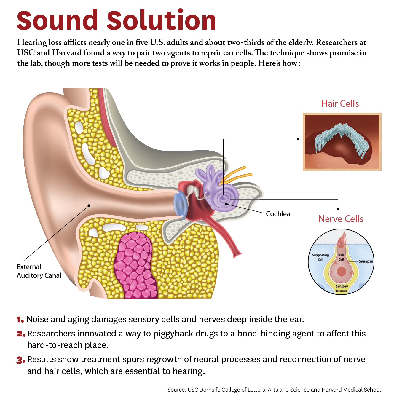
Researchers have developed a new approach to be able to repair cells deep inside the ear. The study, conducted by scientists at University of Southern California (USC) and Harvard University, demonstrates a novel way for a future drug to zero in on damaged nerves and cells inside the ear.

Credit: Matthew Pla Savino/USC News

Credit: Matthew Pla Savino/USC News

“What’s new here is we figured out how to deliver a drug into the inner ear so it actually stays put and does what it’s supposed to do, and that’s novel,” says Charles E. McKenna, Ph.D., a corresponding author for the study and chemistry professor at the USC Dornsife College of Letters, Arts, and Sciences.

“Inside this part of the ear, there’s fluid constantly flowing that would sweep dissolved drugs away, but our new approach addresses that problem. This is a first for hearing loss and the ear,” McKenna adds. “It’s also important because it may be adaptable for other drugs that need to be applied within the inner ear.”

The paper was published April 4 in the journal Bioconjugate Chemistry. The authors include lead researcher Judith S. Kempfle, Ph.D., a 2011 and 2012 Emerging Research Grants scientist, as well as Hearing Restoration Project member Albert Edge, Ph.D., both at Harvard Medical School and The Eaton-Peabody Laboratories in Boston.

There are caveats. The research was conducted on animal tissues in a petri dish. It has not yet been tested in living animals or humans. Yet the researchers are hopeful given the similarities of cells and mechanisms involved. McKenna says since the technique works in the laboratory, the findings provide “strong preliminary evidence” it could work in living creatures. They are already planning the next phase involving animals and hearing loss.

The study breaks new ground because researchers developed a novel drug-delivery method. Specifically, it targets the cochlea, a snail-like structure in the inner ear where sensitive cells convey sound to the brain. Hearing loss occurs due to aging or exposure to noise at unsafe levels. Over time, hair-like sensory cells and bundles of neurons that transmit their vibrations break down, as do ribbon-like synapses, which connect the cells.

The researchers designed a molecule combining 7,8-dihydroxyflavone, which mimics a protein critical for development and function of the nervous system, and bisphosphonate, a type of drug that sticks to bones. This pairing delivered the breakthrough solution, the researchers say, as neurons responded to the molecule and regenerated synapses in mouse ear tissue. This led to the repair of the hair cells and neurons, which are essential to hearing.

“We’re not saying it’s a cure for hearing loss,” McKenna says. “It’s a proof of principle for a new approach that’s extremely promising. It’s an important step that offers a lot of hope.” Hearing loss affects two thirds of people over 70 years and 17 percent of all adults in the United States, and it is expected to nearly double in 40 years.

This is adapted from "Hearing Loss Study at USC, Harvard Shows Hope for Millions" on the USC News website. The authors of the April 4, 2018, Bioconjugate Chemistry study, “Bisphosphonate-Linked TrkB Agonist: Cochlea-Targeted Delivery of a Neurotrophic Agent as a Strategy for the Treatment of Hearing Loss,” include lead researcher Judith S. Kempfle, as well as Christine Hamadani, Nicholas Koen, Albert S. Edge, and David H. Jung of Harvard Medical School and The Eaton-Peabody Laboratories/Massachusetts Eye and Ear in Boston. Kempfle is also affiliated with the University of Tübingen Medical Center. Corresponding author Charles E. McKenna, as well as Kim Nguyen and Boris A. Kashemirov, are at USC Dornsife.

The research was supported by the American Academy of Otolaryngology–Head and Neck Surgery Herbert Silverstein Otology and Neurotology Research Award, an American Otological Society Research Grant, and a grant from the National Institute of Deafness and other Communicative Disorders (R01 DC007174).

Print Friendly and PDF

BLOG ARCHIVE

Sound Processing in Early Brain Regions

To better identify and treat these central auditory processing disorders that appear despite normal ear function, 2016 Emerging Research Grants (ERG) scientist Richard A. Felix II, Ph.D., and colleagues have been investigating how the brain processes complex sounds such as speech.

Print Friendly and PDF

BLOG ARCHIVE

A Clinical Trial for a New Drug to Protect Hearing

By Yishane Lee

The U.S. Food and Drug Administration (FDA) has approved a novel drug to protect against ototoxicity (harmfulness to hearing) due to the use of aminoglycoside antibiotics to treat severe infections. The FDA approval paves the way for a Phase I clinical trial to test whether the drug, found to be significantly protective in animals, is safe for humans.

Mature lateral line hair cells from larval zebrafish (shown with the neuromast sensory organ enlarged) serve as a platform for studying drugs and genes that modulate hair cell susceptibility to ototoxic agents.

Mature lateral line hair cells from larval zebrafish (shown with the neuromast sensory organ enlarged) serve as a platform for studying drugs and genes that modulate hair cell susceptibility to ototoxic agents.

The drug, ORC-13661, was developed by University of Washington professors Edwin Rubel, Ph.D., and David Raible, Ph.D., who are members of Hearing Health Foundation’s Scientific Advisory Board and Hearing Restoration Project, respectively, and Fred Hutchinson Cancer Research Center scientist Julian Simons, Ph.D. “While this program was not directly funded by HHF, both David and I have definitely been supported by HHF for a long time,” Rubel says. “This is a drug to prevent hearing loss that we've developed over the past 15-plus years.”

Rubel points out the drug’s two main features: “It is a brand new drug with a composition of matter patent, not one that is used for other medical purposes and being repurposed; and it is the first drug that was developed, from the get-go, to protect hair cells from ototoxic injury.”

After screening libraries of potential chemicals to see which stopped hair cell death in zebrafish lateral line system, Rubel, Raible, and team identified the best candidate and then boosted its effectiveness by tweaking its chemical structure; results were published in the Journal of Medicinal Chemistry in January 2018.

Rubel adds, “Toxicity studies in zebrafish, rats, and dogs required by the FDA show superior safety and nearly 100 percent hearing protection at all frequencies.” If the Phase I trial shows the drug is safe for humans, the next step is to test its efficacy among patients using aminoglycosides.


Print Friendly and PDF

BLOG ARCHIVE