
A new study suggests that variants of a single gene may alter inner ear development decades before symptoms begin.

Why I Got Surgery (Twice) for My Ménière’s
It has now been 13 years since my vestibular neurectomy, and it ended up being the best decision I ever made for my Ménière’s. I’m back to a somewhat normal life, although I’ve lost about 80 percent of the hearing in my right ear and have high pitched tinnitus in the same ear 24/7.
Understanding Inner Ear Fluid Buildup in Ménière’s Disease
Fluid buildup in the saccule and cochlear duct might be due to increased pressure, while the utricle might be better protected due to its thicker walls and functioning valve. This points to an inverse relationship between membrane thickness and fluid buildup, helping us better understand how fluid buildup occurs in Ménière's disease.
CT Imaging as a Diagnostic Tool for Ménière’s Disease
Comparing the angle measurements of the ATVA, we confirmed the results of the cadaveric study. There was a strong correlation between late onset Ménière’s with a typical “adult” course of the vestibular aqueduct, while early onset Ménière’s was associated with a more straight, “fetal” course of the vestibular aqueduct.
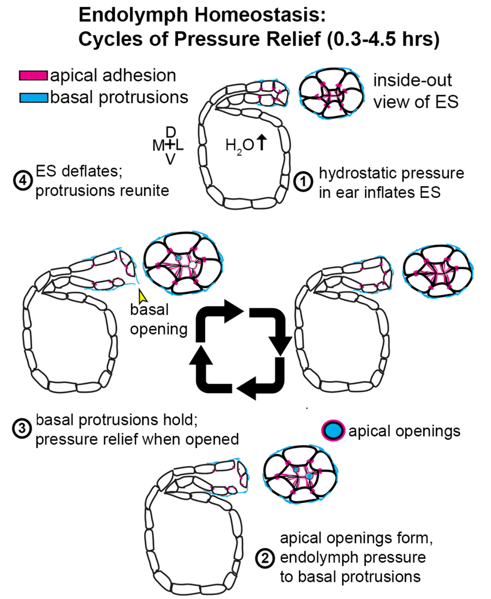
Understanding a Pressure Relief Valve in the Inner Ear
By Ian Swinburne, Ph.D.
The inner ear senses sound to order to hear as well as sensing head movements in order to balance. Sounds or body movements create waves in the fluid within the ear. Specialized cells called hair cells, because of their thin hairlike projections, are submerged within this fluid. Hair cells bend in response to these waves, with channels that open in response to the bending. The makeup of the ear’s internal fluid is critical because as it flows through these channels its contents encode the information that becomes a biochemical and then a neural signal. The endolymphatic sac of the inner ear is thought to have important roles in stabilizing this fluid that is necessary for sensing sound and balance.
This study helps unravel how a valve in the inner ear's endolymphatic sac acts to relieve fluid pressure, one key to understanding disorders affected by pressure abnormalities such as Ménière’s disease.
While imaging transparent zebrafish, my team and I found a pressure-sensitive relief valve in the endolymphatic sac that periodically opens to release excess fluid, thus preventing the tearing of tissue. In our paper published in the journal eLife June 19, 2018, we describe how the relief valve is composed of physical barriers that open in response to pressure. The barriers consist of cells adhering to one another and thin overlapping cell projections that are continuously remodeling and periodically separating in response to pressure.
The unexpected discovery of a physical relief valve in the ear emphasizes the need for further study into how organs control fluid pressure, volume, flow, and ion homeostasis (balance of ions) in development and disease. It suggests a new mechanism underlying several hearing and balance disorders characterized by pressure abnormalities, including Ménière’s disease.

Here is a time-lapse video of the endolymphatic sac, with the sac labeled “pressure relief valve” at 0:40.
2017 Ménière’s Disease Grants scientist Ian A. Swinburne, Ph.D., is conducting research at Harvard Medical School. He was also a 2013 Emerging Research Grants recipient.