
By Steven Barad, M.D.
The cochlea is notoriously difficult to study in live humans—the tiny, snail-like sensory organ buried in a dense skull bone called the temporal bone.
A promising technology is OCT, or optical coherence tomography, which uses laser light to create high resolution images. It’s not unlike ultrasound, but it uses light instead of sound waves.
In August 2026, I visited John Oghalai, M.D., at his University of Southern California laboratory to see this technology in action. Oghalai is developing a compact clinical device based on OCT called the cochleoscope.
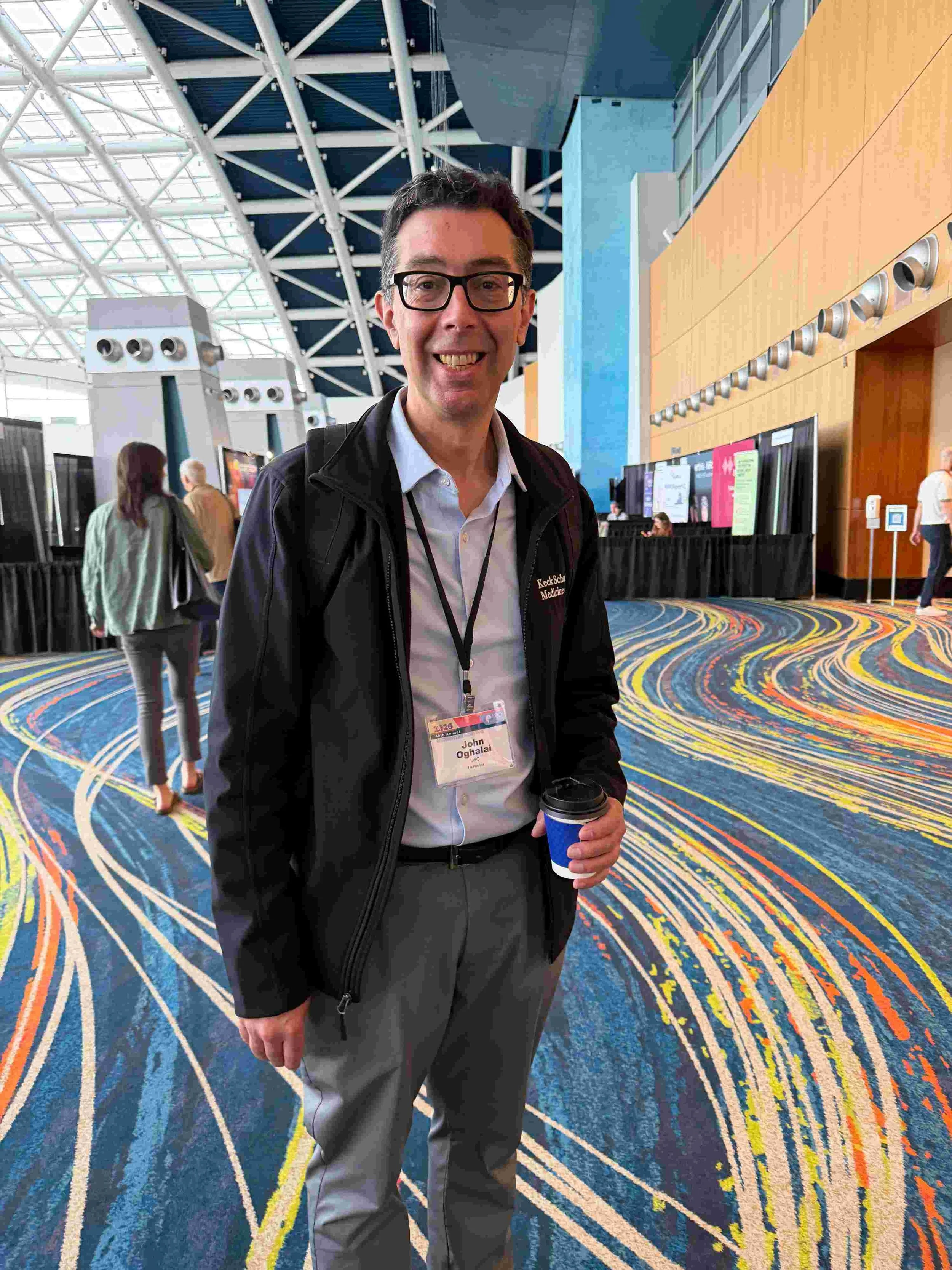
John Oghalai, M.D., at the 2026 Association for Research in Otolaryngology (ARO) MidWinter Meeting.
“You’re basically sending a low-power laser light into tissue,” Oghalai says. “Some of that gets absorbed by the tissue and some of it reflects back. We can detect the reflected light, which gives us a depth profile image.”
Fifteen years ago, he and his colleague, Brian Applegate, Ph.D., began experimenting on mice, and were encouraged by how well OCT worked.
“We could see right through the bone to see almost the whole mouse cochlea and all of the internal structures,” Oghalai says. From there, the team systematically expanded their scope: moving to gerbils, guinea pigs, chickens, and eventually pig temporal bones and chinchillas.
Encouraged by these animal models, the researchers set their sights on clinical translation. In 2025, Oghalai received a National Institutes of Health R01 grant for his project “OCT Imaging of the Human Inner Ear.” The group also received USC funding.
But the bone surrounding the cochlea is thicker in humans than in laboratory animals, so it’s harder for light to penetrate. The more tissue there is to look through, the fuzzier the image becomes.
This barrier introduces a fundamental trade-off in optical design. “You can manipulate light with lenses to get really high resolution,” Oghalai says. “A low power lens gives you a long working distance, but the resolution is not as good. You can see structures, but you can’t see single cells. If we use a higher power lens, the light comes in very steeply, and we can see individual nerve fibers in our animal specimens.”
He adds, “The nice thing about light is that you send in a beam, it travels through a lens, and it comes to a focal point at one spot. So we’re trying to figure out the right tradeoff for what we want to be seeing.”
Currently, the cochleoscope can capture a third to a half of the human cochlea, but not the whole cochlea, and the images are blurry. “Even the thin eardrum distorts the image a little bit,” Oghalai says. “If a patient had a large perforation of their eardrum, we would actually get a better view of the cochlea.”
A Window Into Ear Conditions
Unlike conventional otology research, which relies heavily on scanning electron microscopy of donated cadaver cochleae, OCT promises to capture functional biology in real time.
That visibility could prove revolutionary for complex, poorly understood hearing disorders. Take hyperacusis, a condition where everyday sounds feel unbearably loud, excruciatingly painful, or both. Researchers are exploring whether loudness and pain hyperacusis constitute two separate disorders or one disorder that exists on a continuum of severity.
“I think hyperacusis has a lot to do with inner ear damage, and I’d like to be able to measure that,” Oghalai says. “We just have to see through the thicker bone that humans have in order to make the measurements that define the causes of hearing loss, hyperacusis, and tinnitus.” He and his team are working on this.
Beyond static imaging, the cochleoscope captures how internal structures physically move in response to sound. “We can measure how sounds make the tissues vibrate, which is, of course, critical to hearing,” Oghalai says. “The unique pattern in which they vibrate tells us where the pathology is. We can measure vibrations at a high resolution—basically down to the size of a proton.”
This motion tracking capability offers promise for conditions like Ménière's disease, a balance and hearing condition. “The thought is that Ménière's patients get a buildup of endolymph fluid in their inner ear and it stretches some of the membranes,” he says.
“With the cochleoscope, you can actually watch those membranes stretch, peek into the balance canals, and quantify fluid distention,” Oghalai says. While this can currently be done in the operating room on anesthetized patients, the ultimate goal is routine diagnostic use.
Once the cochleoscope is perfected, “if somebody comes into the clinic with vertigo, we could immediately just take a picture of the inside of their ear and see if it is Ménière's disease or not.”
When the cochleoscope produces clear OCT images, doctors should be able to see what might be wrong inside the ear that causes various hearing conditions. “It could be the hair cells or nerves that are lost, it could be that some of the supporting cells aren’t functioning correctly, or that the blood flow is less than it needs to be,” Oghalai says.
Yet diagnostic precision means little if the imaging process itself poses a threat to sensitive ears. MRI technology—which can detect some inner ear abnormalities but not noise-induced hyperacusis or tinnitus—is loud enough to cause permanent acoustic injury, making it unsuitable for patients with hyperacusis or tinnitus (ringing in the ears). By contrast, Oghalai says, the cochleoscope is non-invasive, and makes only a noise from a fan on the laser.
At this point, the cochleoscope is still a research tool. Oghalai and Applegate are working to make the software more user-friendly, along with scaling down the size and price so that it can be used in clinics more widely. At some point, they will apply for approval from the Food and Drug Administration.
“I’m not sure if OCT is going to be the end-all be-all,” Oghalai says. “But I think it’s a big first step and it’s safe, which is a huge benefit over anything else.”
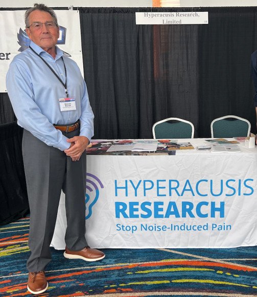
Steven Barad, M.D. (pictured also at ARO), is the president of Hyperacusis Research.
John Oghalai, M.D., is the chair of the USC Caruso Department of Otolaryngology–Head and Neck Surgery, and professor of otolaryngology–head and neck surgery, neurological surgery and biomedical engineering at the Keck School of Medicine of USC and USC Viterbi School of Engineering. He was also a 1996–1997 Emerging Research Grants scientist.
Oghalai and team published their paper “Human inner ear fluid imbalance detected by optical coherence tomography correlates with hearing loss” in Science Translational Medicine in July 2025, also summarized by the Keck School of Medicine. In June 2026, the team’s startup AO Technologies secured up to $3.7 million from ARPA-H to develop the cochleoscope.
Individuals can donate their ears, diseased or healthy, to science by signing up for the National Hearing and Balance Organ Donor Program, formerly the Temporal Bone Registry, which expanded on a program established by Hearing Health Foundation, then called Deafness Research Foundation.
The Latest Blog Posts



The promise of a compact clinical tool capable of imaging deep cochlear tissues through dense bone.