By Lauren McGrath
Hearing Health Foundation (HHF)’s scientists study sensory cells in various species to better understand how they are damaged and how they can be regenerated to restore human hearing.
This quest is the premise of our upcoming Operation Regrow campaign. Your donation will be generously doubled by anonymous donors.
Below are five of the most breathtaking images from our scientists’ labs showcasing the beauty of the hearing and balance functions.

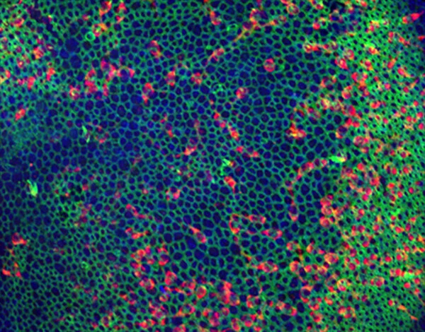
A transverse section through the middle of a post-hatch day 7 chicken cochlea shows tall hair cells in green (detecting Cxcl14 RNA) and short hair cells in red (detecting C14Orf180 RNA). Cell nuclei in blue. Credit: Amanda Janesick, Ph.D./Stanford Otolaryngology.

Confocal microscope image of a Zebrabow fish depicting lateral line neuromasts and ionocytes. Credit: Piotrowski Lab, Stowers Institute for Medical Research.

Single-cell RNA sequencing helped scientists map how sensory hair cells (pink) develop in a newborn mouse cochlea. Credit: Helen Maunsell, NIDCD/NIH
A view of the mouse utricle, a balance organ in the ear. Researchers are working to stimulate complete regeneration in mice. Credit: Jennifer Stone, Ph.D./University of Washington.

Transverse section through the middle of a red-eared slider turtle cochlea shows neuronal connections to hair cells in red-yellow and green, hair cell bundles in white, and cell nuclei in blue. Credit: Amanda Janesick, Ph.D./Stanford Otolaryngology.
New in Hearing and Balance Research


Cochlear implantation (CI) has become an increasingly common audiological solution for children with the most significant degree of unilateral hearing loss—that is, single-sided deafness (SSD)—however, highly variable outcomes are being observed with alarming rates of device nonuse.