By Nesrine Benkafadar, Ph.D.
Losing either type of cochlear sensory hair cells leads to hearing impairment. Inner hair cells convert sound vibrations into electrical signals for the brain to interpret, while outer hair cells amplify sounds to make them easier to detect.
The Hearing Restoration Project has been working to regenerate hair cells. While they regenerate in other species like birds and fish, they do not in mammals, including humans, and once hair cells are damaged or lost, they do not grow back, leading to permanent hearing loss.
A challenge in studying hair cell regeneration has been creating consistent and reliable ways to damage hair cells in laboratory mice. Established methods such as using aminoglycoside antibiotics yield inconsistent and variable hair cell death in mice.
Overcoming this limitation, and as published in Scientific Reports in July 2024, we developed a method involving the surgical delivery of a sisomicin antibiotic solution directly into a specific part of the inner ear, the posterior semicircular canal, in adult mice.
We found this method results in a more predictable and complete loss of hair cells, with the demise of outer hair cells within 14 hours, leading to irreversible hearing loss. Inner hair cells remained until three days post-treatment, after which deterioration in structure and number was observed, culminating in complete hair cell loss by day seven.
The combination of sisomicin and hyperosmotic stress caused consistent and synergistic damage to the hair cells. This means that the high concentration of the antibiotic plus the hyperosmotic condition synergistically worked to eliminate hair cells, making this method of hair cell death more uniform and effective.
These images of a middle region of the mouse cochlear duct show rapid outer hair cell death after sisomicin treatment. (A) Three hours after treatment, outer hair cells appeared normal. (B) Seven hours after treatment, outer hair cells showed signs of death, but inner hair cells and supporting cells are unaffected. (C) Fourteen hours after treatment, all outer hair cells are gone, with inner hair cells and supporting cells still present. Credit: Maraslioglu-Sperber et al./Scientific Reports
Moreover, this method has an applied advantage: its synchronicity and the uniformity of its impact across the entire cochlea. As we have previously shown for the avian inner ear, we expect that this synchronicity, and especially the two distinct waves of hair cell loss, will be useful for determining gene expression changes in dying hair cells and surviving supporting cells.
This robust animal model provides a valuable tool for hair cell regeneration research, facilitating single-cell and omics-based studies toward exploring therapeutic strategies, eventually for humans.
Nesrine Benkafadar, Ph.D., is an instructor in the lab of Hearing Restoration Project member Stefan Heller, Ph.D., who is a professor of otolaryngology–head & neck surgery at Stanford University and a 2001–2002 Emerging Research Grants scientist.


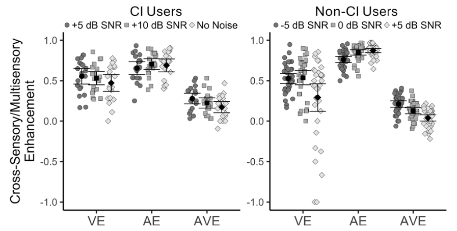
For individuals with long-term hearing loss or severely degraded auditory input, the lack of reliable auditory feedback represents a challenge many orders of magnitude greater than the temporary masking used in this study.