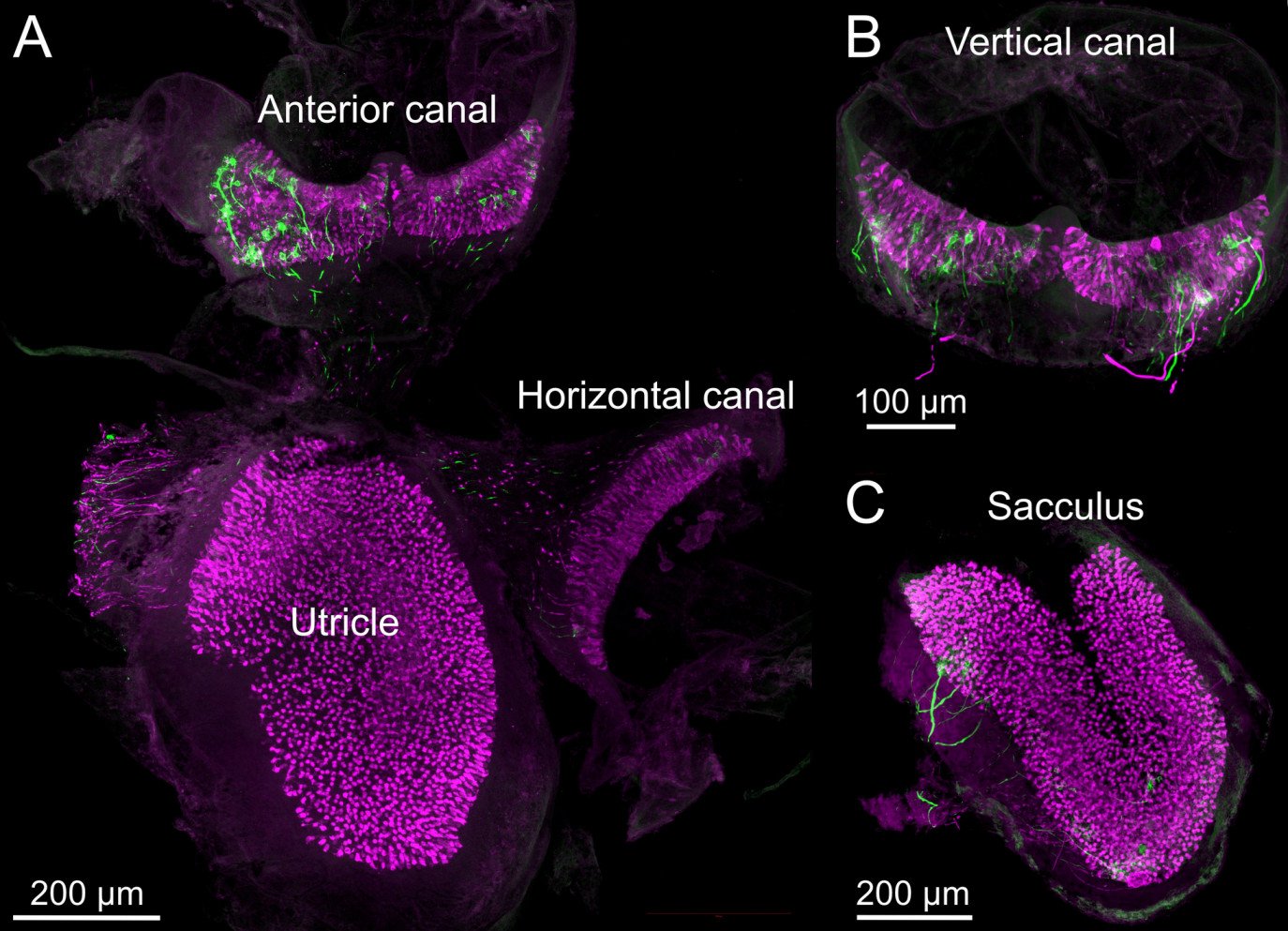
Using the mouse model, vestibular end organ whole mounts imaged with 25× objective. The utricular macula, anterior canal crista, and horizontal canal crista can be dissected out of the temporal bone in one piece. Calretinin (magenta) is expressed in Type I hair cells. Single nerve fibers are labeled with green fluorescent protein. Credit: Balmer and Trussell/Bio-Protocol
The vestibular (balance) sensory apparatus contained in the inner ear is a marvelous evolutionary adaptation for sensing movement in three dimensions and is essential for an animal’s sense of orientation in space, head movement, and balance. Damage to these systems through injury or disease can lead to vertigo, Ménière’s disease, and other disorders that are profoundly debilitating.
One challenge in studying vestibular organs is their location within the bony inner ear and their small size, especially in mice, which have become an advantageous mammalian model. This protocol, published in Bio-Protocol in May 2022, describes the dissection procedure of the five vestibular organs from the inner ear of adult mice.
Immunohistochemical labeling can be used to identify specific hair cell types, such as calretinin-expressing Type I hair cells, or to localize proteins involved in mechanotransduction, cell structure, or vestibular disorders. We then describe the process of immunolabeling of a whole mount preparation using antibodies to label endogenous proteins or to amplify genetically expressed fluorescent proteins for confocal microscopic imaging.
Using typical lab equipment and reagents, a patient technician, student, or postdoc can learn to dissect and immunolabel mouse vestibular organs to investigate their structure in health and disease.
This is adapted from Timothy Balmer, Ph.D., and Laurence Trussell, Ph.D.’s May 2022 Bio-Protocol paper. A 2017 and 2022 ERG scientist, Balmer (left, top) is an assistant professor in the School of Life Sciences at Arizona State University. His 2017 grant was generously funded by the Les Paul Foundation. A 1991 ERG scientist, Trussell is a professor of otolaryngology–head and neck surgery at Oregon Health & Sciences University. Balmer and Trussell employed this procedure in their e-Life study published in April 2019.


Our mission to fund innovative, groundbreaking hearing and balance science is only possible because of you. We are grateful for the support of our community.