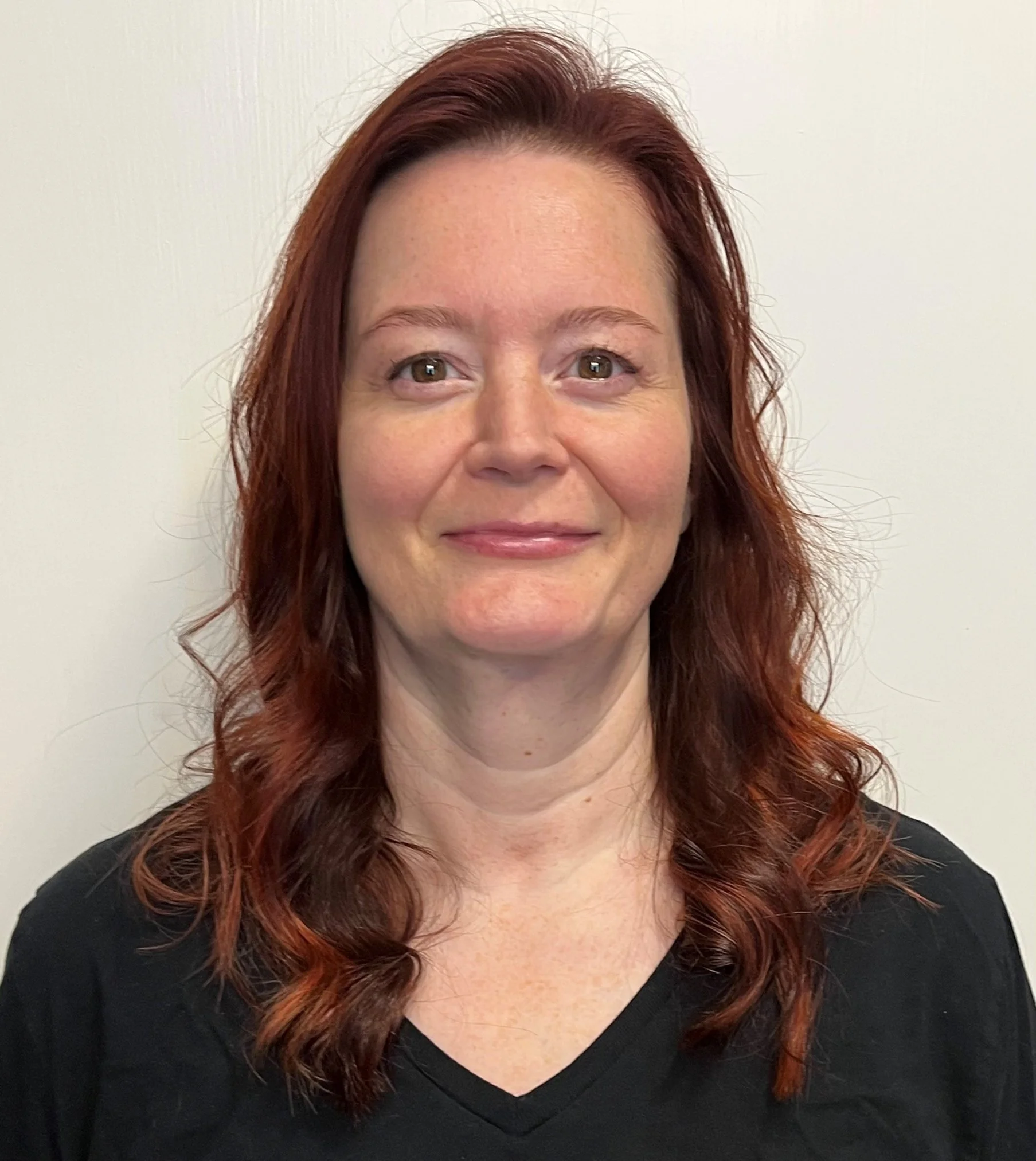
Vijaya Prakash Krishnan Muthaiah, Ph.D.

Vijaya Prakash Krishnan Muthaiah, Ph.D.

University at Buffalo, the State University of New York
Potential of inhibition of poly ADP-ribose polymerase as a therapeutic approach in blast-induced cochlear and brain injury

Many potential drugs in the preclinical phase for treating different types of noise-induced hearing loss (from blast and non-blast noise) revolve around targeting oxidative stress or interfering in the cell death cascade. Though noise-induced oxidative stress and cell death is well studied in the auditory periphery, the effects of noise exposure on the central auditory system remains understudied, especially in blast noise exposure where both auditory and non-auditory structures in the brain are affected. Impulsive noise (blast wave)-induced hearing loss is different from continuous noise exposure as it is more likely to be accompanied by accelerated cognitive deficits, depression, anxiety, dementia, and brain atrophy. It is well established that poly ADP-ribose polymerase (PARP) is a key mediator of cell death and it is overactivated by oxidative stress. Thus this project will explore the potential of PARP inhibition as a potential therapeutic approach for blast-induced cochlear and brain injury. The dampening of PARP overactivation by its inhibitor 3-aminobenzamide is expected to both mitigate blast noise-induced oxidative stress and to interfere with the cell death cascade, thereby reducing cell death in both the peripheral and central auditory system.

Gowri Nayak, Ph.D.

Gowri Nayak, Ph.D.

Cincinnati Children’s Hospital Medical Center
Signaling defects due to Tricellulin deficiency

Mutations in a protein called Tricellulin lead to hereditary hearing loss in humans and degeneration of cochlear sensory cells and deafness in mice. In the inner ear, Tricellulin is found in the sensory epithelia at sites where three epithelial cells meet. The localization of Tricellulin at the junction between cells gives it the potential to respond to external cues and transmit the signals to the cell interior. The current project aims at uncovering the potential cellular signaling roles of Tricellulin by determining the gene expression changes in the inner ear of Tricellulin mutant mice compared to wild-type mice. The results of this study will not only add to the existing knowledge of the inner ear development and maintenance but also will examine the direct effects of losing a protein that helps preserve our hearing.

Research area: Fundamental Auditory Research

Long-term goal of research: To determine the role of cell junction proteins in the inner ear function and elucidate the biological processes that are affected by genetic mutations in these proteins. As tight junctions are also the focus of drug delivery studies, it is valuable to realize the cellular functions of the associated proteins so that they can be manipulated for therapeutic purposes.

Tenzin Ngodup, Ph.D.

Tenzin Ngodup, Ph.D.

Oregon Health & Science University
Discovery of novel inhibitory cell types in the cochlear nucleus

Excessive neuronal electrical activity, or hyperactivity, is believed to underlie tinnitus. While many studies on hyperactivity have focused on a region called the dorsal cochlear nucleus, an auditory processing region in the brainstem, very little attention has been given to the ventral cochlear nucleus (VCN). This is surprising since the VCN is likely required for the activation of higher auditory centers in the brain. One likely cause of hyperexcitability is an imbalance between excitatory and inhibitory neuronal connections, or synapses. With the use of genetically modified mouse lines, we are able to reveal that the diversity of inhibitory cell types and circuitry within the VCN is far richer than previously described. Our primary goal is to discover and study the functional significance of these novel inhibitory neurons in the VCN whose inhibitory action, if compromised, could lead to hyperactivity and auditory dysfunction.

Kirill Vadimovich Nourski, M.D., Ph.D.

Kirill Vadimovich Nourski, M.D., Ph.D.

University of Iowa
Temporal Processing in the Human Auditory Cortex

My research area is the function of the auditory cortex—the hearing center in the brain. Some neurosurgical patients undergo an operation in which arrays of electrodes are temporarily implanted in the brain for clinical diagnostic purposes. This provides a unique opportunity to study how the auditory cortex works, by measuring its activity (“brain waves”) directly from the brain. My personal project involves measuring the brain’s responses to the timing information of sounds and the ability of the brain to accurately follow this timing and use this information to build a coherent percept of the environment. I want to understand where and how, specifically, timing cues are processed in the auditory cortex. Patients with cochlear implants are largely dependent on timing and rhythm cues to understand speech and communicate. On the other hand, people who have auditory processing disorders, may be impaired in their ability to process that kind of information. In order to come up with new ways of assisting people with auditory processing disorders, it’s important to understand how the timing and rhythm of speech is usually handled by the brain.

Research area: fundamental auditory research

Long-term goal of research: Beyond this study, my long term goal is to better understand how the different areas that comprise the auditory cortex in humans are organized and what specific roles they play in processing information about sounds, particularly, as it relates to perception of speech. Ultimately, this knowledge will contribute to finding new and/or improved solutions for people with hearing loss and auditory processing disorders.

Kevin K. Ohlemiller, Ph.D.

Kevin K. Ohlemiller, Ph.D.

Washington University
Cellular and Genetic Bases of Age-Associated Strial Degeneration and EP decline in NOD congenic mice

The electric currents that run through cochlear sensory cells are largely driven by a specialized cochlear structure called the stria vascularis. The work of the stria requires a lot of energy, so that it is densely vascularized (hence the name). Loss of strial blood vessels is thought to be a common cause of age-related hearing loss. Not everyone shows signs of this kind of pathology, however, so that there must be forms of certain genes carried by some people that act as ‘risk’ genes. People who carry ‘risk’ genes may be more likely to experience loss of strial blood vessels, and ultimately loss of the stria itself. In 2008 we discovered that a particular breed of mice (NOD mice) start out with a normal stria, but then show loss of strial vessels, followed by loss of the stria beginning from both ends of the cochlea and progressing toward the middle. These changes were accompanied by other distinctive anatomic features that may tell us something about the process, or may be unrelated. By crossing these mice with another strain that does not show pathology, we will be able to determine what pathologic features are inherited together (thus caused by the same genes), how many genes are involved, and their approximate locations. Any gene(s) we find may have human counterparts that exert similar effects.

Research areas: auditory physiology/pathophysiology, cell biology of hearing and deafness

Long-term goal of research: Finding ‘risk’ genes may not point directly to cures or allow us to predict who will lose their hearing. Nevertheless, identifying the genes, gene networks, and gene products will help pinpoint key reactions that can be tweaked pharmacologically. We are among the first to seek out mouse strains with pathology of the stria vascularis and to use these to uncover genes that promote strial degeneration in mice, and possibly in humans.

Peihan Orestes, Ph.D.

Peihan Orestes, Ph.D.

University of California, Los Angeles
Cellular modifications of the vestibular labyrinth: Intrinsic mechanisms following unilateral aminoglycoside treatment for Meniere’s disease

Intra-tympanic gentamicin is a widely used treatment for unilateral Meniere’s disease, primarily to reduce the activity in the affected ear and thereby reduce the frequency and severity of vertigo attacks. Though the treatments are applied to only one ear, the adaptive effects in the untreated ear have not been widely studied. At the same time, there is evidence indicating that the vestibular receptors exhibit intrinsic capabilities for modification or adaptation to alterations from the normal operating conditions. For example, the inner ear vestibular receptors may undergo a form of “sensory learning” when exposed to changes in the ambient operating conditions associated with spaceflight. An investigation of the contralateral labyrinth following treatments comparable to that associated with intra-tympanic gentamicin may provide clues as to alterations in the conserved ear. These capabilities may be recruited through rehabilitative measures (pharmacologic, physical therapy) to accelerate recovery to normal vestibular function. We propose to study the activity of individual neurons projecting to the conserved vestibular receptors, thereby providing a direct measure of the output of these neurons and by comparisons to our large database of untreated specimens, as well as to determine whether alterations in this activity ensues following adaptation to administration of gentamicin to the contralateral ear.

Research area: Meniere's disease; Ototoxicity

Long-term goal of research: The proposed work is based on the intra-tympanic gentamicin treatment currently used for Ménière’s disease, but the implications of this study could have an even greater impact. Better understanding of the vestibular system’s ability to respond to damage reveals the possibility of retraining the non-lesioned ear, akin to physical therapy. Though most animal studies use unilateral labyrinthectomy as their disease model, such complete loss is rare in the clinical setting. The intra-tympanic gentamicin treatment for Ménière’s disease is not the same as a labyrinthectomy, as the lesions are likely to be partial. Thus, the experiment described here, a direct test of whether peripheral plasticity ensues following partial lesions, is more translationally realistic compared to the labyrinthectomy. Therefore, an investigation into the effects of a less severe lesion, such as the gentamicin regimen proposed here which preserves spontaneous neuronal activity, would be of significant translational value.

Carolyn P. Ojano-Dirain, Ph.D.

Carolyn P. Ojano-Dirain, Ph.D.

University of Florida College of Medicine
Prevention of aminoglycoside-induced hearing loss with the mitochondria-targeted antioxidant MitoQ

Aminoglycoside antibiotics, such as gentamicin, are commonly used to treat serious infections due to bacteria. However, these drugs can cause hearing loss. At present, there is no solution to prevent hearing loss caused by aminoglycoside antibiotics. This research will determine if the antioxidant MitoQ will prevent hearing loss induced by aminoglycoside antibiotics.

Research area: Hearing loss, Ototoxicity

Long-term goal of research: To develop and apply practicable intervention strategies to prevent hearing loss induced by drugs that are toxic to the inner ear.

Melissa Papesh, Au.D., Ph.D.

Melissa Papesh, Au.D., Ph.D.

Portland VA Research Foundation

Central auditory processing disorder and insomnia

At least 26 million American adults complain of hearing and communication difficulties that negatively impact their quality of life despite having no signs of hearing loss. This condition is often referred to auditory processing disorder (APD) reflecting altered processing of auditory information in the brain. While traumatic brain injuries are the most widely known cause for APD, a host of other health conditions are also likely to significantly impact how the brain interprets sound. Our goal with the current project is to examine possible associations between APD and another common condition: insomnia. The objectives of this project are a). to compare peripheral and central auditory system function in patients with normal hearing sensitivity with and without diagnoses of chronic insomnia, and b). to examine the potential for cognitive behavioral therapy (CBT) sleep therapy to improve auditory function in patients with chronic insomnia. In addition to standard hearing tests, we will also measure responses on questionnaires to gauge self-perceived hearing difficulty, assess participants’ ability to discriminate and identify several different types of auditory stimuli, and measure brain responses to sound at multiple levels of the auditory pathway within the brain. Because chronic insomnia is associated with higher rates of cognitive impairment and mental health conditions, we will also measure cognitive function and symptoms of depression and anxiety. Auditory, cognitive and mental health measures will be obtained in patients with diagnosed chronic insomnia both before and after completion of CBT, as well as in a group of control participants following a similar testing timeline. We hypothesize that patients with chronic insomnia will perform more poorly on clinical measures of auditory processing and will report higher rates of hearing handicap compared to controls. In addition, we suspect that that those with chronic insomnia will display abnormally high levels of activity in the brain as well as poor pre-attentive filtering of auditory information compared to good sleepers due to persistent neuronal hyperarousal. Finally, our hope is that these auditory manifestations will improve once participants complete CBT thus providing a pathway to improved hearing in a subset of patients experiencing APD.

Patrick D. Parker, Ph.D.

Patrick D. Parker, Ph.D.

Johns Hopkins University School of Medicine

Emergence of tonotopically organized spontaneous activity in the brain after genetic disruption of MET channel

Secondary disorders develop alongside hearing loss, such as the perception of phantom sounds (tinnitus) and a hypersensitivity to sound (hyperacusis). These disorders have no curative treatment, in part due to our poor understanding of the underlying neurobiology. Using experimental models of hearing loss, I recently discovered that sound-independent (SI) patterns of neural activity emerge in the brain’s auditory centers that resemble those elicited by sound in hearing mice. This finding indicates that the brain can self- generate neural patterns that resemble sound processing, independent of the inner ear. To understand the relevance of SI activity to human disorders like tinnitus, I’ll expand these findings to more translationally relevant models of hearing loss (loud noise exposure and age-related hearing loss), as well as determine the area of the brain that generates these patterns. The results of these studies may help to develop new approaches to treat tinnitus, hyperacusis, and related disorders.

Christian N. Paxton, Ph.D.

Christian N. Paxton, Ph.D.

University of Utah

The role of Fgf4 in otic placode induction

Development and patterning of the inner ear is a complex process that is mediated by several signaling molecules, including members of the fibroblast growth factor (FGF) family. We recently found that Fgf4 is expressed in the ear-forming region just prior to the induction of ear development. Fgf4 has not previously been described in the induction or formation of the inner ear. Based on its temporal and spatial pattern of expression we hypothesize that Fgf4 is involved in the early processes of ear development and propose to investigate its role(s) in these processes by determining whether it is sufficient and/or required to induce the early stages of inner ear development. We also will examine the signals responsible for localizing Fgf4 expression to the otic forming domain.

Z. Ellen Peng, Ph.D.

Z. Ellen Peng, Ph.D.

University of Wisconsin-Madison
Investigating cortical processing during comprehension of reverberant speech in adolescents and young adults with cochlear implants

Through early cochlear implant (CI) fitting, many children diagnosed with profound neurosensorial hearing loss gain access to verbal communications through electrical hearing and go on to develop spoken language. Despite good speech outcomes tested in sound booths, many children experience difficulties in understanding speech in most noisy and reverberant indoor environments. While up to 75 percent of their time learning is spent in classrooms, the difficulty from adverse acoustics adding to children’s processing of degraded speech from CI is not well understood. In this project, we examine speech understanding in classroom-like environments through immersive acoustic virtual reality. In addition to behavioral responses, we measure neural activity using functional near-infrared spectroscopy (fNIRS)—a noninvasive, CI-compatible neuroimaging technique, in cortical regions that are responsible for speech understanding and sustained attention. Our findings will reveal the neural signature of speech processing by CI users, who developed language through electrical hearing, in classroom-like environments with adverse room acoustics.

Tatjana Piotrowski, Ph.D.

Tatjana Piotrowski, Ph.D.

University of Utah Medical School

Molecular analysis of hair cell regeneration in the zebrafish lateral line

We are aiming to elucidate the genetic pathways underlying hair cell regeneration in zebrafish with the long-term goal of activating these pathways in mammals. Our lab is taking a twofold approach to identify genes involved in hair cell regeneration. We are performing gene expression analyses from mantle cells of control larvae and from larvae in which mantle cells are proliferating to regenerate killed hair cells (as proposed in this application). As a second approach we are performing a mutagenesis screen for zebrafish mutants which are not able to regenerate hair cells, and thus carry mutations in regeneration-specific genes. A prominent cause of deafness is loss of hair cells due to age, noise or antibiotic treatments. In contrast to mammalian hair cells, fish, bird and amphibian hair cells turn over frequently and regenerate following hair cell death. Little is known why lower vertebrates are able to regenerate hair cells but humans do not. This is partly due to the relative inaccessibility of inner ear hair cells to direct observation and manipulation. Our aim is to take advantage of the lateral line of zebrafish to define and characterize the molecular and cellular interactions occurring during hair cell regeneration. If successful, our results will set the stage for testing whether hair cell regeneration can be activated in humans.

Sarah F. Poissant, Ph.D.

Sarah F. Poissant, Ph.D.

University of Massachusetts, Amherst
The Impact of Total Communication on the Auditory Perception of Speech

For decades, most children with severe-to-profound hearing loss were educated in special schools for the deaf. In more recent years, increasing numbers of these children have been partially or fully main-streamed and educated along-side their peers with normal hearing. Much debate has ensued regarding the best language of instruction (sign-only, sign+speech, speech only) for them. It is generally thought that a symbolic, gesture-based language system, such as manually-coded English used as part of simultaneous communication methods, provides a facilitative benefit. However, there is not enough information about how children combine manual and spoken cues in this type of communication system to draw firm conclusions about optimal approaches to classroom teaching that best support aural reception of spoken language. We plan to ask and answer a very specific question: What is the direct effect of simultaneously delivered sign language on the perception of speech for children with hearing loss developing spoken language? The research approach builds from the observation that perception of speech that has been artificially degraded (e.g., to mimic a hearing loss) is strikingly improved when listeners have knowledge of the content of the message. The proposed study applies this hypothesis to children with hearing loss to determine whether signs serve in part as a prime to improve auditory perception of speech.

Research area: Auditory Development; Congenital Hearing Loss; Fundamental Auditory Research

Long-term goal of research: To assess how total communication – the combined use of manual signs, speech, and speech-reading – can most effectively be employed as a habilitation strategy to improve auditory perceptual abilities.

Melissa Polonenko, Ph.D.

Melissa Polonenko, Ph.D.

University of Minnesota–Twin Cities

Identifying hearing loss through neural responses to engaging stories

Spoken language acquisition in children with hearing loss relies on early identification of hearing loss followed by timely fitting of hearing devices to ensure they receive an adequate representation of the speech that they need to hear. Yet current tests for young children rely on non-speech stimuli, which are processed differently by hearing aids and do not fully capture the complexity of speech. This project will develop a new and efficient test - called multiband peaky speech - that uses engaging narrated stories and records responses from the surface of the head (EEG) to identify frequency-specific hearing loss. Computer modeling and EEG experiments in adults will determine the best combination of parameters and stories to speed up the testing for use in children, and evaluate the test’s ability to identify hearing loss. This work lays the necessary groundwork for extending this method to children and paves the way for clinics to use this test as a hearing screener for young children–and ultimately our ability to provide timely, enhanced information to support spoken language development.

The long-term goal is to develop an engaging, objective clinical test that uses stories to identify hearing loss in young children and evaluate changes to their responses to the same speech through hearing aids. This goal addresses two important needs identified by the U.S.’s Early Hearing Detection and Intervention (EHDI) program, and will positively impact the developmental trajectory of thousands of children who need monitoring of their hearing status and evaluation of outcomes with their hearing devices.

Generously funded by Royal Arch Research Assistance

Erin K. Purcell, Ph.D.

Erin K. Purcell, Ph.D.

Sonja Pyott, Ph.D.

Sonja Pyott, Ph.D.

University of North Carolina Wilmington

Enhancement of the efferent-hair cell synapse by metabotropic glutamate receptors

This proposal aims to improve our understanding of the molecular mechanisms regulating synapses in the cochlea and will specifically characterize how a class of molecules, metabotropic glutamate receptors (mGluRs), regulates the efferent-hair cell synapses. Sensory hair cells of the cochlea communicate with the brain at specialized sites called synapses. Inner hair cells have numerous afferent synapses that relay information about sound from the hair cell to the brain. In contrast, outer hair cells are characterized by efferent synapses from the brain that regulate hair cell activity. Although these efferent and afferent synapses are normally considered to be independent from one another, experiments studying immature inner hair cells suggest that glutamate, the neurotransmitter required for transmission at the afferent synapse, may also modify the response of the efferent synapse. Efferent innervation of the cochlea is thought to protect against noise-induced hearing loss. Considering that noise-induced hearing loss accounts for one-third of all cases of deafness, understanding the mechanisms regulating efferent synapses is of special clinical relevance. This project will investigate this hypothesis and should uncover novel pharmaceutical targets to modulate the efferent synaptic response to either dampen hair cell activity and prevent noise-induced hearing or boost hair cell activity and combat deafness.

Kelly Radziwon, Ph.D.

Kelly Radziwon, Ph.D.

State University of New York at Buffalo
Noise-induced hyperacusis in rats with and without hearing loss

Hyperacusis is an auditory perceptual disorder in which everyday sounds are perceived as uncomfortably or excruciatingly loud. Researchers and audiologists assess hyperacusis in the clinic by asking patients to rate sounds based on their perceived loudness, resulting in a measure known as a loudness discomfort level (LDL). Loudness discomfort ratings are a useful clinical tool, but in the lab we cannot ask animals to “rate” sounds. Instead, to measure loudness perception in animals, our lab trains rats to detect a variety of sounds of varying intensity. By measuring how quickly the animals respond to each sound—faster in reaction to higher intensity sounds and more slowly to lower intensity sounds—we can obtain an accurate picture of perceived loudness in animals. By comparing electrophysiological recordings with behavioral performances of the individual animals, this project aims to characterize the relationship between changes in neural activity and loudness perception in animals with and without noise-induced hearing loss.

The relationship between pain-associated proteins in the auditory pathway and hyperacusis

Hyperacusis is a condition in which sounds of moderate intensity are perceived as intolerably loud or even painful. Despite the apparent link between pain and hyperacusis in humans, little research has been conducted directly comparing the presence of inflammation along the auditory pathway and the occurrence of hyperacusis. One of the major factors limiting this research has been the lack of a reliable animal behavioral model of hyperacusis. However, using reaction time measurements as a marker for loudness perception, I have successfully assessed rats for drug-induced hyperacusis and, more recently, noise-induced hyperacusis. Briefly, the animals will be trained to detect noise bursts of varying intensity. As in humans, the rats will respond faster with increasing sound intensity. Following drug administration or noise exposure, rats will be deemed to have hyperacusis if they have faster-than-normal reaction times to moderate and high-level sounds. Therefore, the goal of the proposed research is to correlate the presence of pain-related molecules along the auditory pathway with reliable behavioral measures of drug and noise-induced hyperacusis.

Lavanya Rajagopalan, Ph.D.

Lavanya Rajagopalan, Ph.D.

Baylor College of Medicine

The structural and functional basis of electromotility in prestin, the outer ear amplifier protein

Prestin, a membrane protein in outer hair cells in the cochlea, is involved in cochlear amplification leading to frequency sensitivity. The long-term objectives of this study are to understand the molecular basis of prestin function, to advance the field closer to designing therapeutics in certain types of hearing loss. This will provide insight into the molecular basis of prestin-related hearing loss, and can lead to rational design of therapeutics to treat such conditions.

Robert Raphael, Ph.D.

Robert Raphael, Ph.D.

Rice University
Understanding the biophysics and protein biomarkers of Ménière’s disease via optical coherence tomography imaging

Our sense of hearing and balance depends on maintaining proper fluid balance in a specialized fluid in the inner ear called the endolymph. Ménière’s disease is an inner ear disorder associated with increased fluid pressure in the endolymph that involves dizziness, hearing loss, and tinnitus. Ménière’s disease is difficult to diagnose and treat clinically, which is a source of frustration for both physicians and patients. Part of the barrier to diagnosing and treating Ménière’s disease is the lack of imaging tools to study the inner ear and a poor understanding of the underlying causes. The goal of this research is to develop an approach to noninvasively image the inner ear and study the internal structures in the vestibular system in typical and disease states. We will utilize optical coherence tomography (OCT), a technique capable of imaging through bone, and observe changes in the fluid compartments in the inner ear. The expected outcome of this research will be the establishment of a powerful non-invasive imaging platform of the inner ear that will enable us to test hypotheses, in living animals, on how ion transport regulates the endolymph, how disorders of ion transport cause disruption of endolymphatic fluid, and how the expression of different biomarkers lead to disorders of ion transport.

Khaleel Razak, Ph.D.

 Khaleel Razak, Ph.D.

University of California, Riverside
Age-related hearing loss and cortical processing

Presbycusis (age-related hearing loss) is one of the most prevalent forms of hearing impairment in humans, and contributes to speech recognition impairments and cognitive decline. Both peripheral and central auditory system changes are involved in presbycusis. The relative contributions of peripheral hearing loss and brain aging to presbycusis-related auditory processing declines remain unclear. This project will address this question by comparing genetically engineered, age-matched mice with one group experiencing presbycusis and a second group that does not. Spectrotemporal processing (such as speech processing) will be studied as an outcome measure.