By the National Institute on Deafness and Other Communication Disorders (NIDCD)

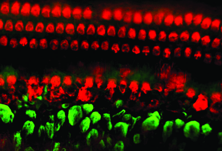
Line of polarity reversal (LPR) and location of Emx2 within two inner ear structures. Arrows indicate hair bundle orientation. Source: eLife
Using animal models, scientists have demonstrated that a protein called Emx2 is critical to how specialized cells that are important for maintaining hearing and balance are positioned in the inner ear. Emx2 is a transcription factor, a type of protein that plays a role in how genes are regulated. Conducted by scientists at the National Institute on Deafness and Other Communication Disorders (NIDCD), part of the National Institutes of Health (NIH), the research offers new insight into how specialized sensory hair cells develop and function, providing opportunities for scientists to explore novel ways to treat hearing loss, balance disorders, and deafness. The results are published March 7, 2017, in eLife.
Our ability to hear and maintain balance relies on thousands of sensory hair cells in various parts of the inner ear. On top of these hair cells are clusters of tiny hair-like extensions called hair bundles. When triggered by sound, head movements, or other input, the hair bundles bend, opening channels that turn on the hair cells and create electrical signals to send information to the brain. These signals carry, for example, sound vibrations so the brain can tell us what we’ve heard or information about how our head is positioned or how it is moving, which the brain uses to help us maintain balance.
NIDCD researchers Doris Wu, Ph.D., chief of the Section on Sensory Cell Regeneration and Development and member of HHF’s Scientific Advisory Board, which provides oversight and guidance to our Hearing Restoration Project (HRP) consortium; Katie Kindt, Ph.D., acting chief of the Section on Sensory Cell Development and Function; and Tao Jiang, a doctoral student at the University of Maryland College Park, sought to describe how the hair cells and hair bundles in the inner ear are formed by exploring the role of Emx2, a protein known to be essential for the development of inner ear structures. They turned first to mice, which have been critical to helping scientists understand how intricate parts of the inner ear function in people.
Each hair bundle in the inner ear bends in only one direction to turn on the hair cell; when the bundle bends in the opposite direction, it is deactivated, or turned off, and the channels that sense vibrations close. Hair bundles in various sensory organs of the inner ear are oriented in a precise pattern. Scientists are just beginning to understand how the hair cells determine in which direction to point their hair bundles so that they perform their jobs.
In the parts of the inner ear where hair cells and their hair bundles convert sound vibrations into signals to the brain, the hair bundles are oriented in the same direction. The same is true for hair bundles involved in some aspects of balance, known as angular acceleration. But for hair cells involved in linear acceleration—or how the head senses the direction of forward and backward movement—the hair bundles divide into two regions that are oriented in opposite directions, which scientists call reversed polarity. The hair bundles face either toward or away from each other, depending on whether they are in the utricle or the saccule, two of the inner ear structures involved in balance. In mammals, the dividing line at which the hair bundles are oriented in opposite directions is called the line of polarity reversal (LPR).
Using gene expression analysis and loss- and gain-of-function analyses in mice that either lacked Emx2 or possessed extra amounts of the protein, the scientists found that Emx2 is expressed on only one side of the LPR. In addition, they discovered that Emx2 reversed hair bundle polarity by 180 degrees, thereby orienting hair bundles in the Emx2 region in opposite directions from hair bundles on the other side of the LPR. When the Emx2 was missing, the hair bundles in the same location were positioned to face the same direction.
Looking to other animals to see if Emx2 played the same role, they found that Emx2 reversed hair bundle orientation in the zebrafish neuromast, the organ where hair cells with reversed polarity that are sensitive to water movement reside.
These results suggest that Emx2 plays a key role in establishing the structural basis of hair bundle polarity and establishing the LPR. If Emx2 is found to function similarly in humans, as expected, the findings could help advance therapies for hearing loss and balance disorders. They could also advance research into understanding the mechanisms underlying sensory hair cell development within organs other than the inner ear.
This work was supported within the intramural laboratories of the NIDCD (ZIA DC000021 and ZIA DC000085).
Doris Wu Ph.D. is member of HHF’s Scientific Advisory Board, which provides oversight and guidance to our Hearing Restoration Project (HRP) consortium This article was repurpsed with permission from the National Institute on Deafness and Other Communication Disorders.