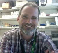
Martin Basch, Ph.D.
Meet the Researcher
Martin Basch, Ph.D. received his Ph.D. in developmental biology at the California Institute of Technology. He worked as a postdoctoral fellow with Drs. Andrew Groves and Neil Segil at House Ear Institute. He is currently an Instructor in Dr. Andrew Groves’ lab at Baylor.
The Research
Baylor College of Medicine
Development of Biomarkers to Study Strial Development and Degeneration
The stria vascularis is a specialized tissue in the inner ear, localized in the lateral wall of the cochlea. This tissue generates endolymph, a special fluid that is rich in potassium and provides the driving force for the function of the sensory cells in the ear. Strial defects are implicated in many human syndromes involving profound hearing loss and are one of the main causes of presbycusis (age related hearing loss). In spite of its importance for normal hearing, we know very little about the development of the stria vascularis. The goal of this project is to identify the genes that are responsible for strial development, and that present the potential to restore or regenerate damaged stria vascularis in cases of both congenital or age related hearing loss.
Live imaging of the developing cochlea
During development the cochlear duct grows from the ventral side of the inner ear and a prosensory domain is established that contains common progenitors for hair cells and supporting cells. As these cells differentiate they form a highly organized pattern that in mammals consists of one row of inner hair cells (IUC) and three rows of outer hair cells (OHC), each surrounded by supporting cells. The organization of this intricate pattern is concomitant with the elongation of the cochlea from the base to the apex. Because there is little or no cell proliferation at this time, these processes rely heavily on cell movements. We propose that an active process of cell movement is responsible for achieving the complex architecture of the cochlear epithelium. In recent years we began to understand the molecular mechanisms underlying hair cell and supporting cell differentiation. However, the cellular mechanisms involved in this process have been difficult to study. To address this question, we have developed an organ culture method that allows us to study the individual movements of genetically labeled cells through 3 dimensional time-lapse confocal microscopy.
Long-Term Goal: To understand how the development of the stria vascularis and apply this knowledge towards the regeneration and/or repair of damaged stria vascularis in cases of congential defects or age related hearing loss.


