By Timothy S. Balmer, Ph.D., and Laurence O Trussell, Ph.D.

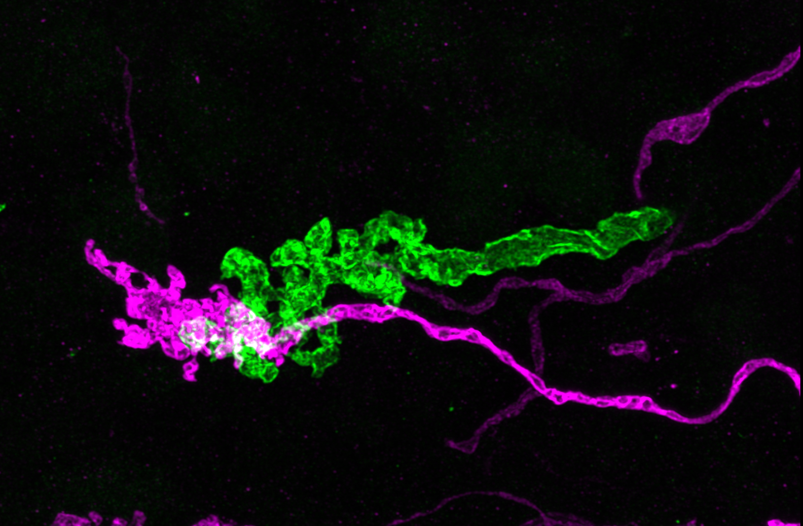
Balmer & Trussell traced the direct and indirect pathways that carry vestibular information to the cerebellum for controlling balance and posture. Shown here is a primary afferent axon (green) expressing the light-gated ion channel, Channelrhodopsin. Postsynaptic cells, in this case a unipolar brush cell (magenta), were recorded from during stimulation of the input axons by light flashes. This technique was used to discover how direct and indirect vestibular pathways are processed in the cerebellum.
Mice are helping scientists to understand how the world around us remains looking stable even as we move.
While out jogging, you have no trouble keeping your eyes fixed on objects in the distance even though your head and eyes are moving with every step. Humans owe this stability of the visual world partly to a region of the brain called the vestibular cerebellum. From its position underneath the rest of the brain, the vestibular cerebellum detects head motion and then triggers compensatory movements to stabilize the head, body and eyes.
The vestibular cerebellum receives sensory input from the body via direct and indirect routes. The direct input comes from five structures within the inner ear, each of which detects movement of the head in one particular direction. The indirect input travels to the cerebellum via the brainstem, which connects the brain with the spinal cord. The indirect input contains information on head movements in multiple directions combined with input from other senses such as vision.
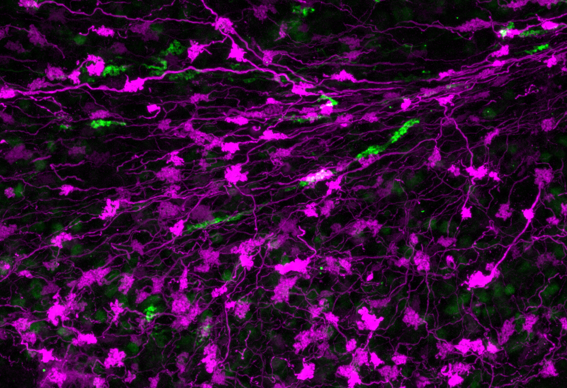
Balmer & Trussell traced the direct and indirect pathways that carry vestibular information to the cerebellum for controlling balance and posture. Direct projections from the vestibular inner ear (green) and indirect projections from the brainstem (magenta) were shown to target different populations of neurons in the cerebellum.


By studying the mouse brain, Balmer and Trussell have now mapped the direct and indirect circuits that carry sensory information to the vestibular cerebellum. Both types of input activate cells within the vestibular cerebellum called unipolar brush cells (UBCs). There are two types of UBCs: ON and OFF. Direct sensory input from the inner ear activates only ON UBCs. These cells respond to the arrival of sensory input by increasing their activity. Indirect input from the brainstem activates both ON UBCs and OFF UBCs. The latter respond to the input by decreasing their activity.

The vestibular cerebellum thus processes direct and indirect inputs via segregated pathways containing different types of UBCs. The next step in understanding how the cerebellum maintains a stable visual world is to identify the circuitry beyond the UBCs. Understanding these circuits will ultimately provide insights into balance disorders, such as vertigo.
A 2017 Emerging Research Grants (ERG) scientist who received the Les Paul Foundation Award for Tinnitus Research, Timothy Balmer, Ph.D., is a postdoctoral fellow at the Oregon Hearing Research Center at Oregon Health & Science University (OHSU). Laurence Trussell, Ph.D., a 1991 ERG recipient, is a professor of otolaryngology–head and neck surgery at OHSU.
This research summary was repurposed with permission from eLife with permission.
