By Ngoc-Nhi Luu, M.D., Dr. med.
Ménière’s disease is an inner ear condition with symptoms including vertigo, hearing loss, and tinnitus, and may be associated with an accumulation of fluid in the inner ear, termed endolymphatic hydrops. Diagnosis of Ménière’s is entirely based on clinical characteristics, and to date, no classification has been established that can predict the onset or course of the disease. Patients with Ménière’s can have varying degrees of symptoms, so defining subtypes within the Ménière’s population may help establish a classification to improve diagnoses and treatments.
Our previous analysis of cadaveric ears of patients with Ménière’s revealed striking differences within the endolymphatic sac in the inner ear, which regulates endolymph fluid. We had found two different aberrations of the endolymphatic sac—its underdevelopment or its degeneration—among Ménière’s patients, suggesting that the loss of endolymphatic sac cell function and the possible impairment of endolymphatic fluid regulation may lead to Ménière’s. In addition, these two pathologies may be associated with differing clinical traits of the disease.
In our prior work, we examined sections of human cadaveric inner ears with Ménière’s and found differences in the angular trajectory of the vestibular aqueduct (ATVA), the bony canal in which the endolymphatic sac is located. These differences resembled either the trajectory of typical adults, or the trajectory of early developmental, fetal vestibular aqueducts. ATVA similar to other adults without Ménière’s were associated with late onset of the condition, whereas Ménière’s patients with “fetal” ATVA experienced early onset.
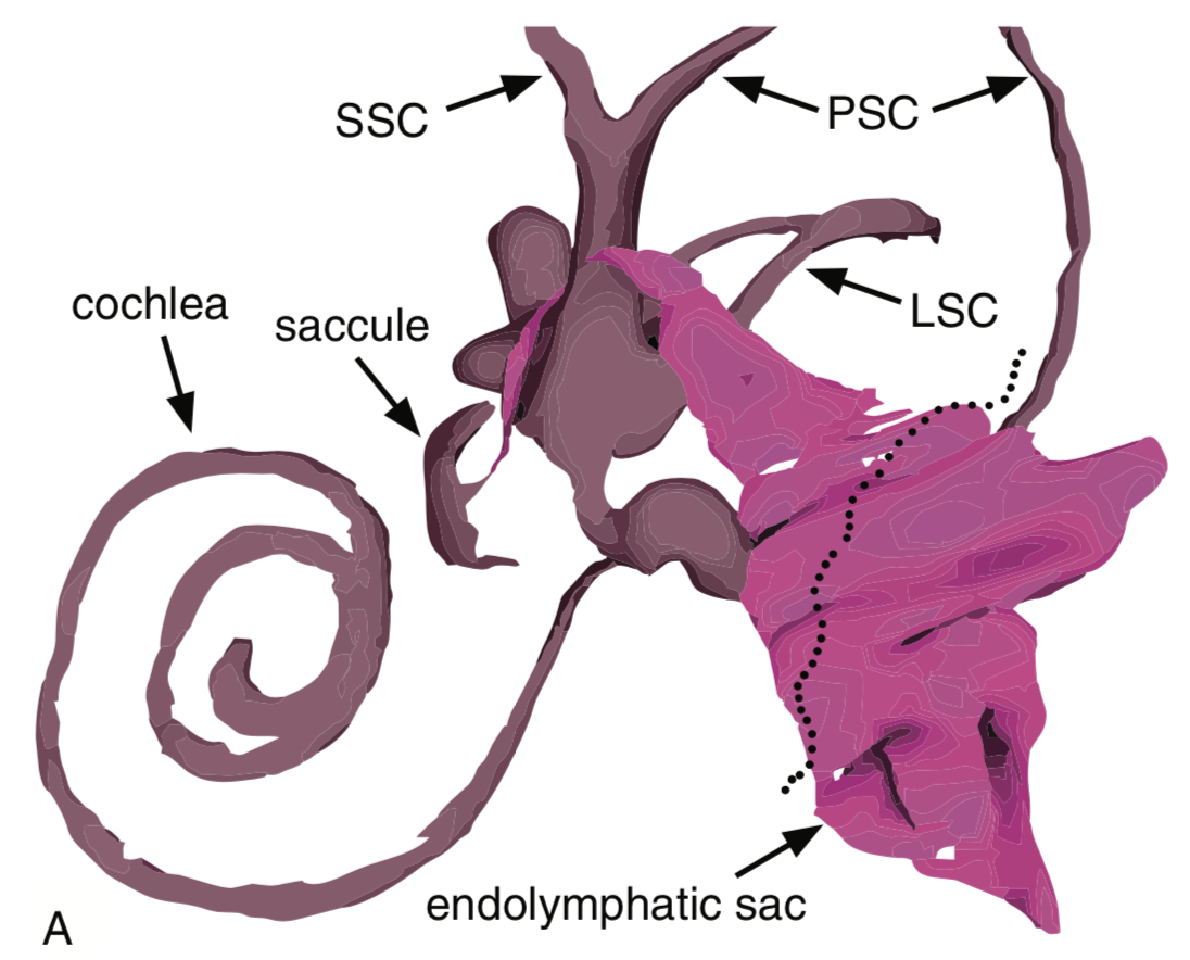
A 3D reconstruction of the endolymphatic space of a typical human adult inner ear. In Ménière’s disease patients, the anatomy of the endolymphatic sac differs, suggesting that the impairment of the sac’s function to regulate fluid may lead to Ménière’s. (LSC, lateral semicircular canal; PSC, posterior semicircular canal; SCC, superior semicircular canal.)
For our paper published in the journal Otology & Neurotology in April 2019, we hypothesized that this difference can be detected with a CT scan (computerized tomography) in patients with early or late onset Ménière’s.
We used a custom-made, open-source web application for angle measurements and applied this technique on high resolution CT imaging of patients with Ménière’s. Comparing the angle measurements of the ATVA, we confirmed the results of the cadaveric study. There was a strong correlation between late onset Ménière’s with a typical “adult” course of the vestibular aqueduct, while early onset Ménière’s was associated with a more straight, “fetal” course of the vestibular aqueduct.
As such our study aims to develop a radiographic screening tool, such as a CT scan of the inner ear, to classify different Ménière’s subtypes. It appears that early onset Ménière’s patients have a different anatomy of the vestibular aqueduct compared with late onset Ménière’s patients.
We want to better understand if these findings also correlate with additional clinical factors, such as specific symptoms or a positive family history for Ménière’s. Ultimately, this may help to further characterize different Ménière’s subtypes in order to better diagnose, predict the course of, and treat the condition.

Ngoc-Nhi Luu, M.D., Dr. med., is a postdoctoral fellow at Eaton-Peabody Laboratories, Massachusetts Eye and Ear, Harvard Medical School, and an ENT resident at University Hospital Zurich. Luu’s 2017 ERG grant was generously funded by The Estate of Howard F. Schum. Coauthors on the paper include Judith Kempfle, M.D. (a 2010 ERG scientist), Steven Rauch, M.D. (1990 ERG), and Joseph Nadol, M.D. (1976–77 ERG).
